1XDM image
Deposition Date 2004-09-07
Release Date 2005-03-22
Last Version Date 2023-08-23
Entry Detail
PDB ID:
1XDM
Keywords:
Title:
Structure of human aldolase B associated with hereditary fructose intolerance (A149P), at 291K
Biological Source:
Source Organism:
Homo sapiens (Taxon ID: 9606)
Host Organism:
Method Details:
Experimental Method:
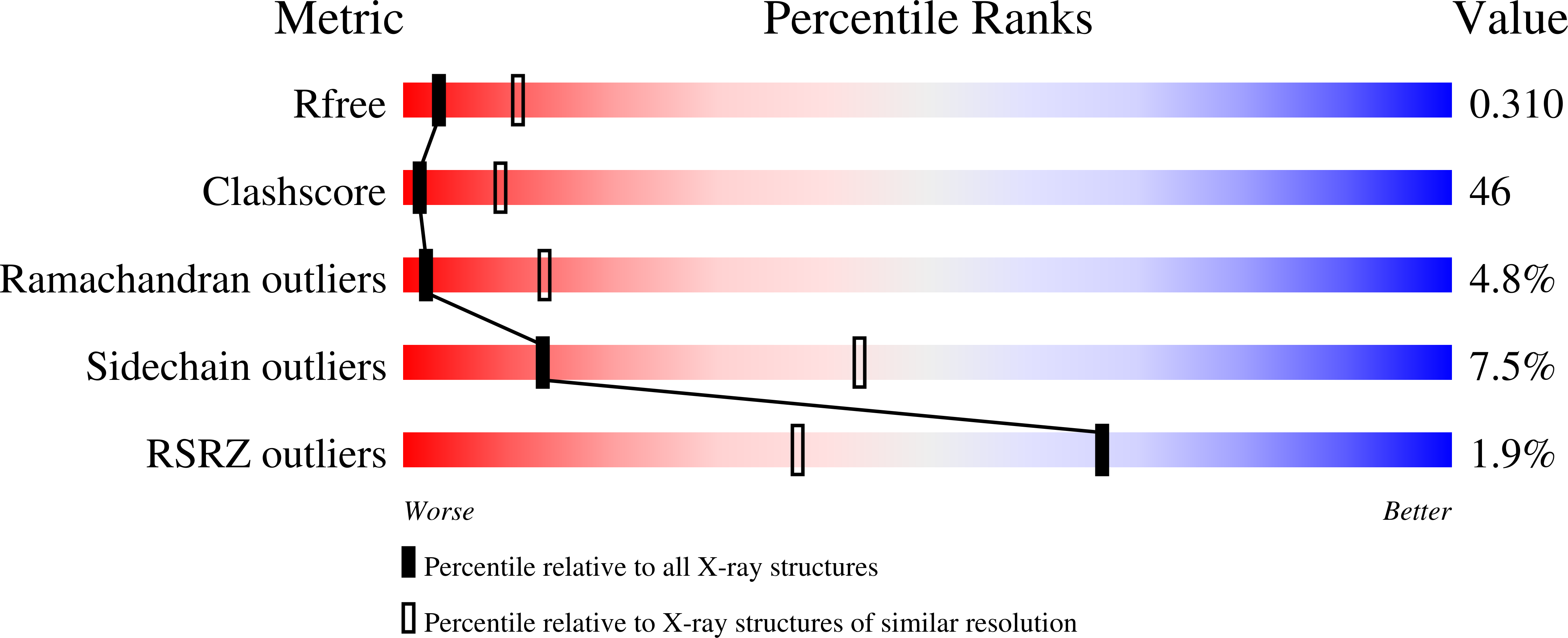
Resolution:
3.00 Å
R-Value Free:
0.31
R-Value Work:
0.27
R-Value Observed:
0.27
Space Group:
P 21 21 2
Macromolecular Entities
Polymer Type:polypeptide(L)
Molecule:Fructose-bisphosphate aldolase B
Gene (Uniprot):ALDOB
Mutations:A149P
Chain IDs:A, B, C, D, E (auth: W), F (auth: X), G (auth: Y), H (auth: Z)
Chain Length:365
Number of Molecules:8
Biological Source:Homo sapiens
Ligand Molecules
Primary Citation
Structure of the thermolabile mutant aldolase B, A149P: molecular basis of hereditary fructose intolerance.
J.Mol.Biol. 347 135 144 (2005)
PMID: 15733923 DOI: 10.1016/j.jmb.2005.01.008

Abstact

Hereditary fructose intolerance (HFI) is a potentially lethal inborn error in metabolism caused by mutations in the aldolase B gene, which is critical for gluconeogenesis and fructose metabolism. The most common mutation, which accounts for 53% of HFI alleles identified worldwide, results in substitution of Pro for Ala at position 149. Structural and functional investigations of human aldolase B with the A149P substitution (AP-aldolase) have shown that the mutation leads to losses in thermal stability, quaternary structure, and activity. X-ray crystallography is used to reveal the structural basis of these perturbations. Crystals of AP-aldolase are grown at two temperatures (4 degrees C and 18 degrees C), and the structure solved to 3.0 angstroms resolution, using the wild-type structure as the phasing model. The structures reveal that the single residue substitution, A149P, causes molecular disorder around the site of mutation (residues 148-159), which is propagated to three adjacent beta-strand and loop regions (residues 110-129, 189-199, 235-242). Disorder in the 110-129-loop region, which comprises one subunit-subunit interface, provides an explanation for the disrupted quaternary structure and thermal instability. Greater structural perturbation, particularly at a Glu189-Arg148 salt bridge in the active-site architecture, is observed in the structure determined at 18 degrees C, which could explain the temperature-dependent loss in activity. The disorder revealed in these structures is far greater than that predicted by homology modeling and underscores the difficulties in predicting perturbations of protein structure and function by homology modeling alone. The AP-aldolase structure reveals the molecular basis of a hereditary disease and represents one of only a few structures known for mutant proteins at the root of the thousands of other inherited disorders.

Legend

Protein

Chemical

Disease

Primary Citation of related structures