Deposition Date
2004-09-05
Release Date
2005-03-22
Last Version Date
2024-11-20
Entry Detail
PDB ID:
1XDC
Keywords:
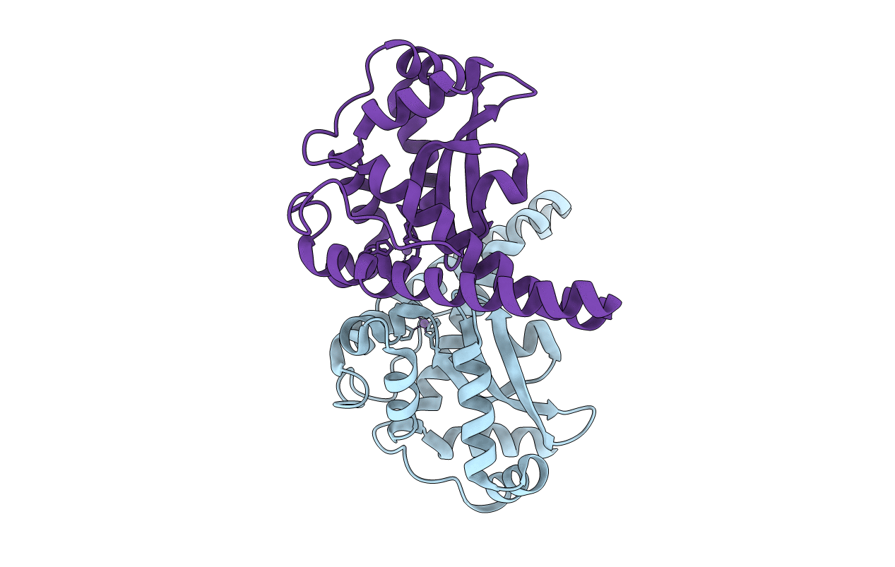
Title:
Hydrogen Bonding in Human Manganese Superoxide Dismutase containing 3-Fluorotyrosine
Biological Source:
Source Organism(s):
Homo sapiens (Taxon ID: 9606)
Expression System(s):
Method Details:
Experimental Method:
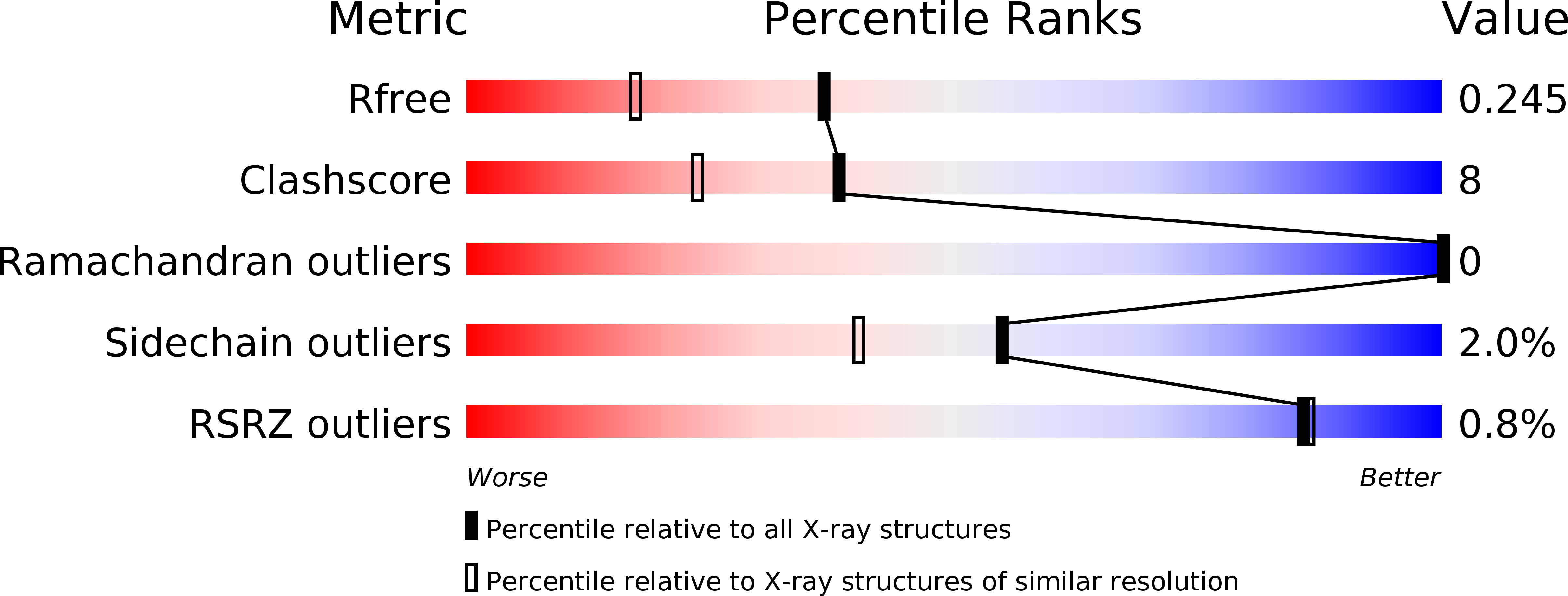
Resolution:
1.85 Å
R-Value Free:
0.25
R-Value Work:
0.21
R-Value Observed:
0.21
Space Group:
P 21 21 2