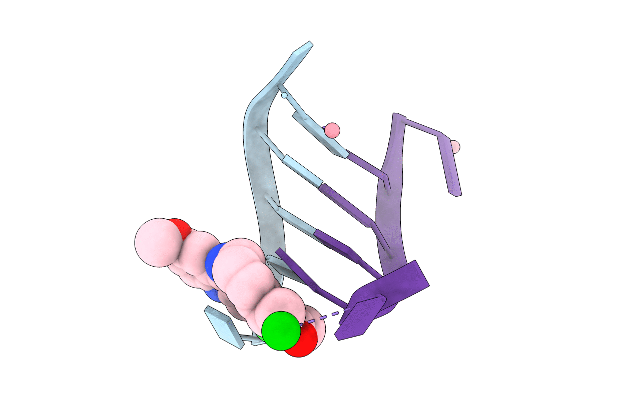
1XCS image
Deposition Date 2004-09-03
Release Date 2005-07-19
Last Version Date 2024-02-14
Entry Detail
PDB ID:
1XCS
Keywords:
Title:
structure of oligonucleotide/drug complex
Biological Source:
Source Organism:
(Taxon ID: ) (Taxon ID: )
Method Details:
Experimental Method:
Resolution:
1.40 Å
R-Value Free:
0.25
R-Value Work:
0.19
R-Value Observed:
0.19
Space Group:
C 2 2 2
Macromolecular Entities
Polymer Type:polydeoxyribonucleotide
Molecule:5'-D(*CP*GP*TP*AP*CP*G)-3'
Chain IDs:A, B
Chain Length:6
Number of Molecules:2
Biological Source:
Primary Citation
Variable role of ions in two drug intercalation complexes of DNA
J.Biol.Inorg.Chem. 10 476 482 (2005)
PMID: 15926069 DOI: 10.1007/s00775-005-0655-3

Abstact

The crystal structures of the hexamer duplex d(CGTACG)(2) complexed with the intercalating anthraquinone derivative 1,5-bis[3-(diethylamino)propionamido]anthracene-9,10-dione and the acridine derivative 9-acridinyl tetralysine have been solved at 2.0- and 1.4-A resolution, respectively. In both cases, the drugs adopt multiple orientations within a large DNA cavity constituted by two groups of four approximately coplanar bases. Cations play a pivotal role in the crystal structure. Both complexes crystallise in the presence of Co(2+), Ba(2+) and Na(+) ions. They reveal at least two different types of coordination environments: (1) specific sites for Co(2+) interacting with N7 of guanine; (2) a central ionic site formed by four phosphate groups, which can be occupied by different ions. One more ionic site that is not always occupied by ions is also visible in the electron density map. All of them play a role in the crystal structure.

Legend

Protein

Chemical

Disease

Primary Citation of related structures