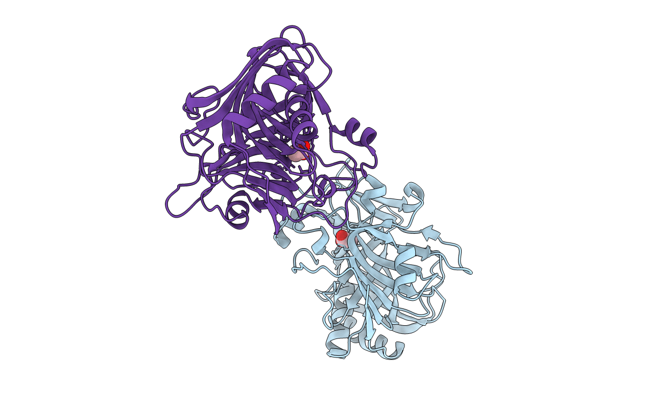
1XCR image
Deposition Date 2004-09-03
Release Date 2005-09-27
Last Version Date 2024-04-03
Entry Detail
PDB ID:
1XCR
Title:
Crystal Structure of Longer Splice Variant of PTD012 from Homo sapiens reveals a novel Zinc-containing fold
Biological Source:
Source Organism:
Homo sapiens (Taxon ID: 9606)
Host Organism:
Method Details:
Experimental Method:
Resolution:
1.70 Å
R-Value Free:
0.22
R-Value Work:
0.18
Space Group:
P 1 21 1
Macromolecular Entities
Polymer Type:polypeptide(L)
Molecule:hypothetical protein PTD012
Gene (Uniprot):C11orf54
Chain IDs:A, B
Chain Length:316
Number of Molecules:2
Biological Source:Homo sapiens
Primary Citation
Crystal structure of Homo sapiens PTD012 reveals a zinc-containing hydrolase fold
Protein Sci. 15 914 920 (2006)
PMID: 16522806 DOI: 10.1110/ps.052037006

Abstact

The human protein PTD012 is the longer product of an alternatively spliced gene and was described to be localized in the nucleus. The X-ray structure analysis at 1.7 A resolution of PTD012 through SAD phasing reveals a monomeric protein and a novel fold. The shorter splice form was also studied and appears to be unfolded and non-functional. The structure of PTD012 displays an alphabetabetaalpha four-layer topology. A metal ion residing between the central beta-sheets is partially coordinated by three histidine residues. X-ray absorption near-edge structure (XANES) analysis identifies the PTD012-bound ion as Zn(2+). Tetrahedral coordination of the ion is completed by the carboxylate oxygen atom of an acetate molecule taken up from the crystallization buffer. The binding of Zn(2+) to PTD012 is reminiscent of zinc-containing enzymes such as carboxypeptidase, carbonic anhydrase, and beta-lactamase. Biochemical assays failed to demonstrate any of these enzyme activities in PTD012. However, PTD012 exhibits ester hydrolase activity on the substrate p-nitrophenyl acetate.

Legend

Protein

Chemical

Disease

Primary Citation of related structures