1XCD image
Entry Detail
PDB ID:
1XCD
Title:
Dimeric bovine tissue-extracted decorin, crystal form 1
Biological Source:
Source Organism:
PDB Version:
Deposition Date:
2004-09-01
Release Date:
2004-11-02
Method Details:
Experimental Method:
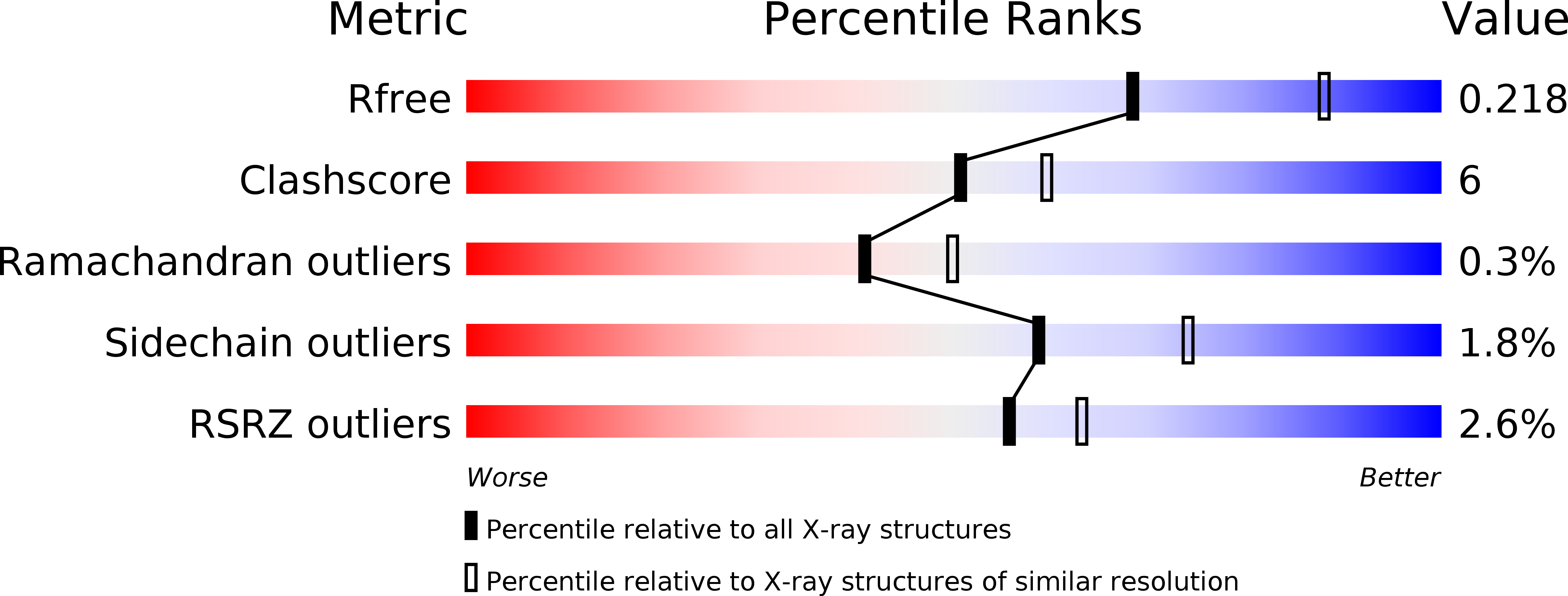
Resolution:
2.31 Å
R-Value Free:
0.25
R-Value Work:
0.22
R-Value Observed:
0.22
Space Group:
C 2 2 21
Macromolecular Entities
Polymer Type:polypeptide(L)
Description:Decorin
Chain IDs:A
Chain Length:329
Number of Molecules:1
Biological Source:Bos taurus
Modified Residue
Compound ID Chain ID Parent Comp ID Details 2D Image
ASN A ASN GLYCOSYLATION SITE
Primary Citation
Crystal structure of the dimeric protein core of decorin, the archetypal small leucine-rich repeat proteoglycan
Proc.Natl.Acad.Sci.Usa 101 15633 15638 (2004)
PMID: 15501918 DOI: 10.1073/pnas.0402976101

Abstact

Decorin is a ubiquitous extracellular matrix proteoglycan with a variety of important biological functions that are mediated by its interactions with extracellular matrix proteins, cytokines, and cell surface receptors. Decorin is the prototype of the family of small leucine-rich repeat proteoglycans and proteins (SLRPs), characterized by a protein core composed of leucine-rich repeats (LRRs), flanked by two cysteine-rich regions. We report here the crystal structure of the dimeric protein core of decorin, the best characterized member of the SLRP family. Each monomer adopts the curved solenoid fold characteristic of LRR domains, with a parallel beta-sheet on the inside interwoven with loops containing short segments of beta-strands, 3(10) helices, and polyproline II helices on the outside. Two main features are unique to this structure. First, decorin dimerizes through the concave surfaces of the LRR domains, which have been implicated previously in protein-ligand interactions. The amount of surface buried in this dimer rivals the buried surfaces of some of the highest-affinity macromolecular complexes reported to date. Second, the C-terminal region adopts an unusual capping motif that involves a laterally extended LRR and a disulfide bond. This motif seems to be unique to SLRPs and has not been observed in any other LRR protein structure to date. Possible implications of these features for decorin ligand binding and SLRP function are discussed.

Legend

Protein

Chemical

Disease

Primary Citation of related structures