Deposition Date
2004-09-01
Release Date
2005-09-06
Last Version Date
2023-08-23
Entry Detail
PDB ID:
1XC8
Keywords:
Title:
CRYSTAL STRUCTURE COMPLEX BETWEEN THE WILD-TYPE LACTOCOCCUS LACTIS FPG (MUTM) AND A FAPY-DG CONTAINING DNA
Biological Source:
Source Organism(s):
Lactococcus lactis subsp. cremoris (Taxon ID: 1359)
Expression System(s):
Method Details:
Experimental Method:
Resolution:
1.95 Å
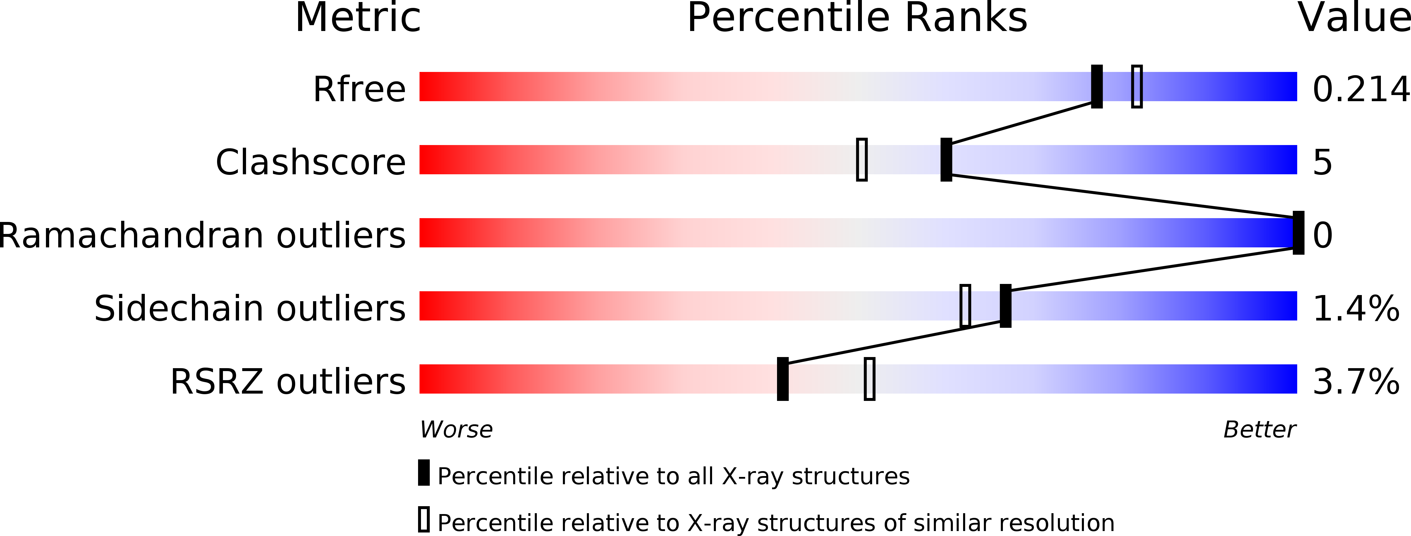
R-Value Free:
0.20
R-Value Work:
0.17
R-Value Observed:
0.17
Space Group:
P 41 21 2