1XAI image
Deposition Date 2004-08-25
Release Date 2005-03-01
Last Version Date 2023-08-23
Entry Detail
PDB ID:
1XAI
Keywords:
Title:
CRYSTAL STRUCTURE OF STAPHLYOCOCCUS AUREUS 3-DEHYDROQUINATE SYNTHASE (DHQS) IN COMPLEX WITH ZN2+, NAD+ AND CARBAPHOSPHONATE
Biological Source:
Source Organism:
Host Organism:
Method Details:
Experimental Method:
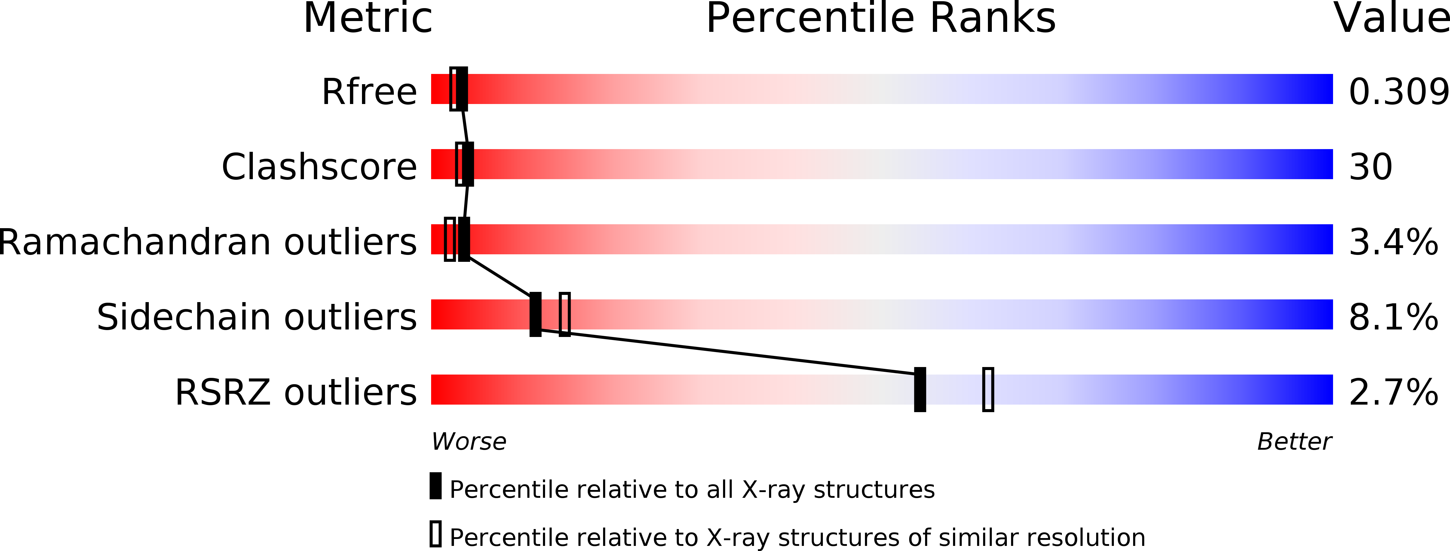
Resolution:
2.30 Å
R-Value Free:
0.31
R-Value Work:
0.21
R-Value Observed:
0.21
Space Group:
P 43
Macromolecular Entities
Structures with similar UniProt ID
Protein Blast
Polymer Type:polypeptide(L)
Molecule:3-dehydroquinate synthase
Gene (Uniprot):aroB
Chain IDs:A, B
Chain Length:354
Number of Molecules:2
Biological Source:Staphylococcus aureus
Primary Citation
Comparison of ligand induced conformational changes and domain closure mechanisms, between prokaryotic and eukaryotic dehydroquinate synthases.
J.Mol.Biol. 343 533 546 (2004)
PMID: 15465043 DOI: 10.1016/j.jmb.2004.08.039

Abstact

Dehydroquinate synthase (DHQS) is a potential target for the development of novel broad-spectrum antimicrobial drugs, active against both prokaryotes and lower eukaryotes. Structures have been reported for Aspergillus nidulans DHQS (AnDHQS) in complexes with a range of ligands. Analysis of these AnDHQS structures showed that a large-scale domain movement occurs during the normal catalytic cycle, with a complex series of structural elements propagating substrate binding-induced conformational changes away from the active site to distal locations. Compared to corresponding fungal enzymes, DHQS from bacterial species are both mono-functional and significantly smaller. We have therefore determined the structure of Staphylococcus aureus DHQS (SaDHQS) in five liganded states, allowing comparison of ligand-induced conformational changes and mechanisms of domain closure between fungal and bacterial enzymes. This comparative analysis shows that substrate binding initiates a large-scale domain closure in both species' DHQS and that the active site stereochemistry, of the catalytically competent closed-form enzyme thus produced, is also highly conserved. However, comparison of AnDHQS and SaDHQS open-form structures, and analysis of the putative dynamic processes by which the transition to the closed-form states are made, shows a far lower degree of similarity, indicating a significant structural divergence. As a result, both the nature of the propagation of conformational change and the mechanical systems involved in this propagation are quite different between the DHQSs from the two species.

Legend

Protein

Chemical

Disease

Primary Citation of related structures
Feedback Form
Name
Email
Institute
Feedback