1X9I image
Deposition Date 2004-08-21
Release Date 2004-12-07
Last Version Date 2023-08-23
Entry Detail
PDB ID:
1X9I
Keywords:
Title:
Crystal structure of Crystal structure of phosphoglucose/phosphomannose phosphoglucose/phosphomannoseisomerase from Pyrobaculum aerophilum in complex with glucose 6-phosphate
Biological Source:
Source Organism:
Host Organism:
Method Details:
Experimental Method:
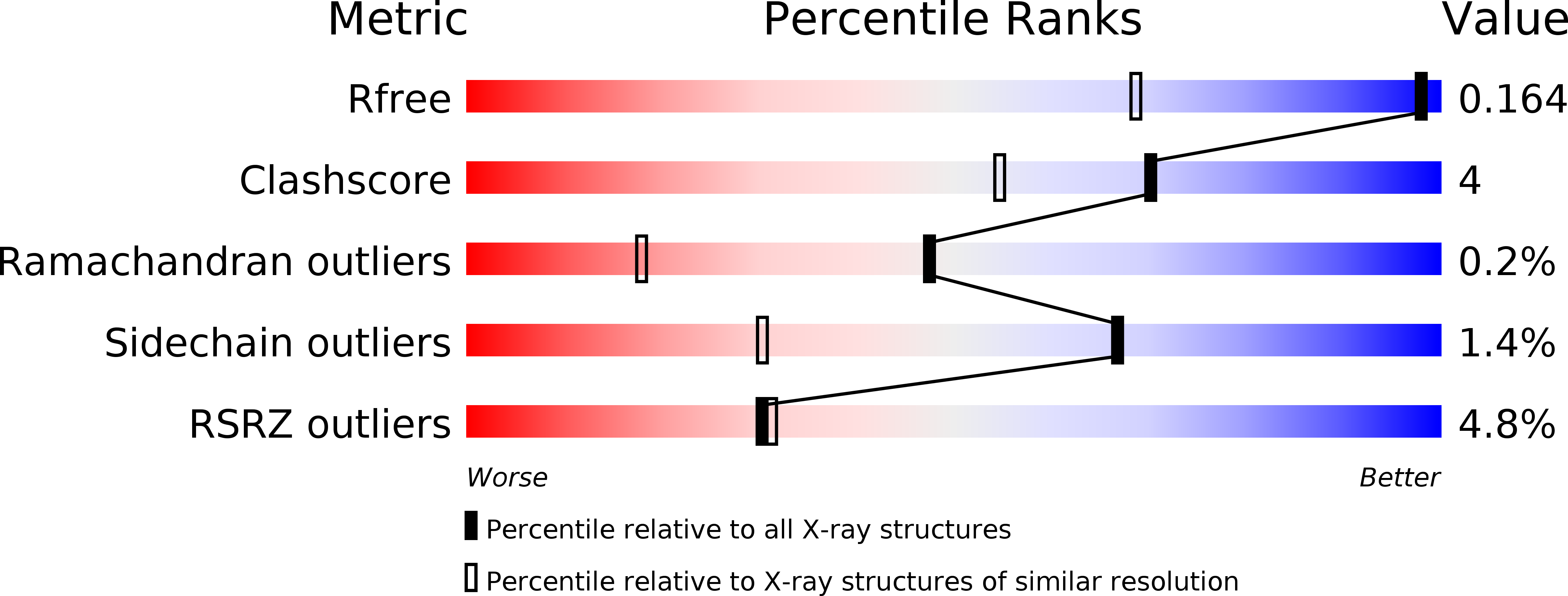
Resolution:
1.16 Å
R-Value Free:
0.16
R-Value Work:
0.14
R-Value Observed:
0.14
Space Group:
P 1 21 1
Macromolecular Entities
Structures with similar UniProt ID
Protein Blast
Polymer Type:polypeptide(L)
Molecule:glucose-6-phosphate isomerase
Chain IDs:A, B
Chain Length:302
Number of Molecules:2
Biological Source:Pyrobaculum aerophilum
Primary Citation
Structural basis for phosphomannose isomerase activity in phosphoglucose isomerase from Pyrobaculum aerophilum: a subtle difference between distantly related enzymes.
Biochemistry 43 14088 14095 (2004)
PMID: 15518558 DOI: 10.1021/bi048608y

Abstact

The crystal structure of a dual-specificity phosphoglucose/phosphomannose isomerase from the crenarchaeon Pyrobaculum aerophilum (PaPGI/PMI) has been determined in complex with glucose 6-phosphate at 1.16 A resolution and with fructose 6-phosphate at 1.5 A resolution. Subsequent modeling of mannose 6-phosphate (M6P) into the active site of the enzyme shows that the PMI activity of this enzyme may be due to the additional space imparted by a threonine. In PGIs from bacterial and eukaryotic sources, which cannot use M6P as a substrate, the equivalent residue is a glutamine. The increased space may permit rotation of the C2-C3 bond in M6P to facilitate abstraction of a proton from C2 by Glu203 and, after a further C2-C3 rotation of the resulting cis-enediolate, re-donation of a proton to C1 by the same residue. A proline residue (in place of a glycine in PGI) may also promote PMI activity by positioning the C1-O1 region of M6P. Thus, the PMI reaction in PaPGI/PMI probably uses a cis-enediol mechanism of catalysis, and this activity appears to arise from a subtle difference in the architecture of the enzyme, compared to bacterial and eukaryotic PGIs.

Legend

Protein

Chemical

Disease

Primary Citation of related structures