Deposition Date
2004-08-20
Release Date
2005-11-22
Last Version Date
2023-08-23
Entry Detail
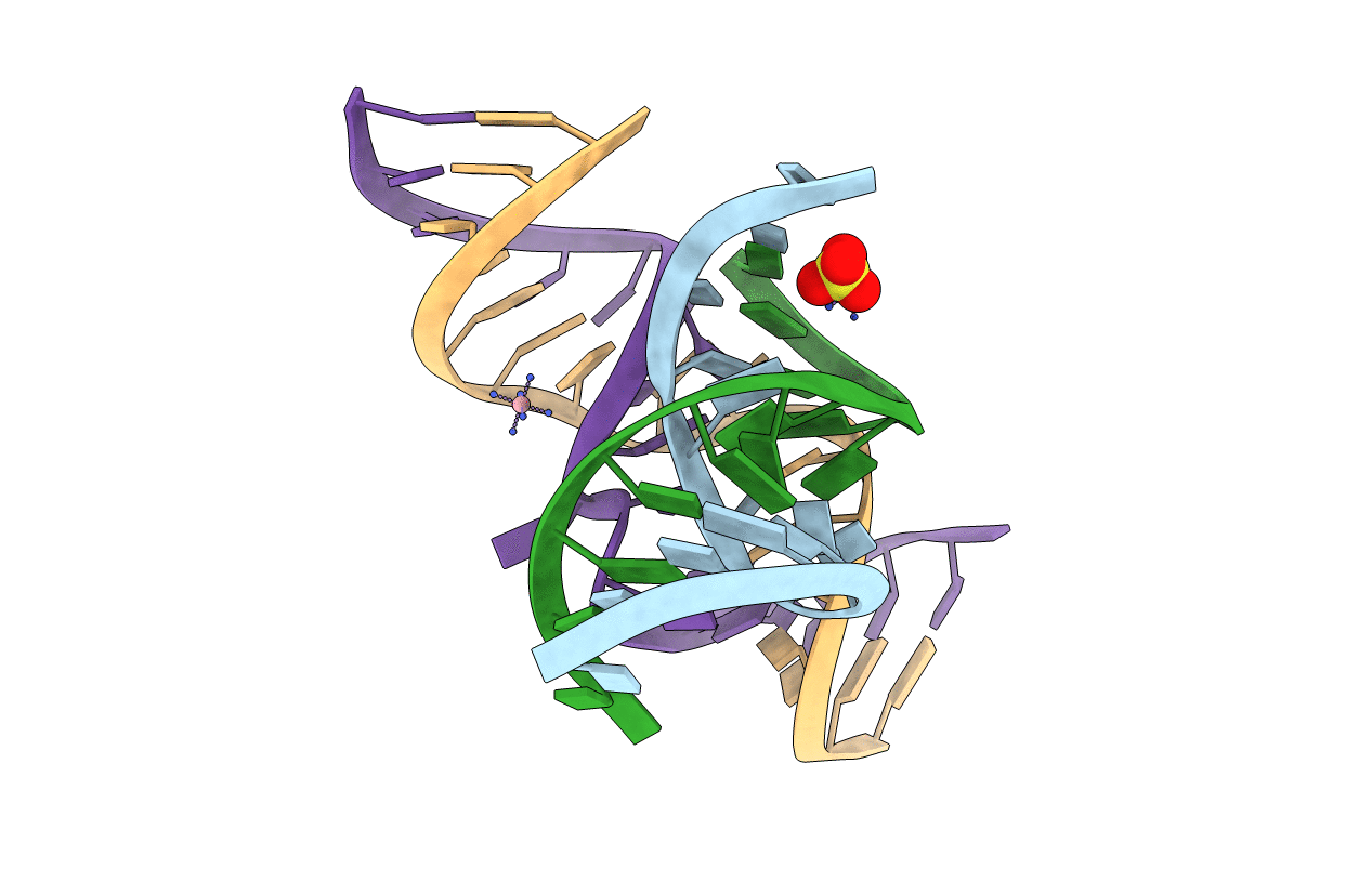
Method Details:
Experimental Method:
Resolution:
2.19 Å
R-Value Free:
0.26
R-Value Work:
0.24
R-Value Observed:
0.24
Space Group:
P 61 2 2