1X8S image
Deposition Date 2004-08-18
Release Date 2004-12-07
Last Version Date 2024-02-14
Entry Detail
PDB ID:
1X8S
Keywords:
Title:
Structure of the Par-6 PDZ domain with a Pals1 internal ligand
Biological Source:
Source Organism:
Drosophila melanogaster (Taxon ID: 7227)
(Taxon ID: )
Method Details:
Experimental Method:
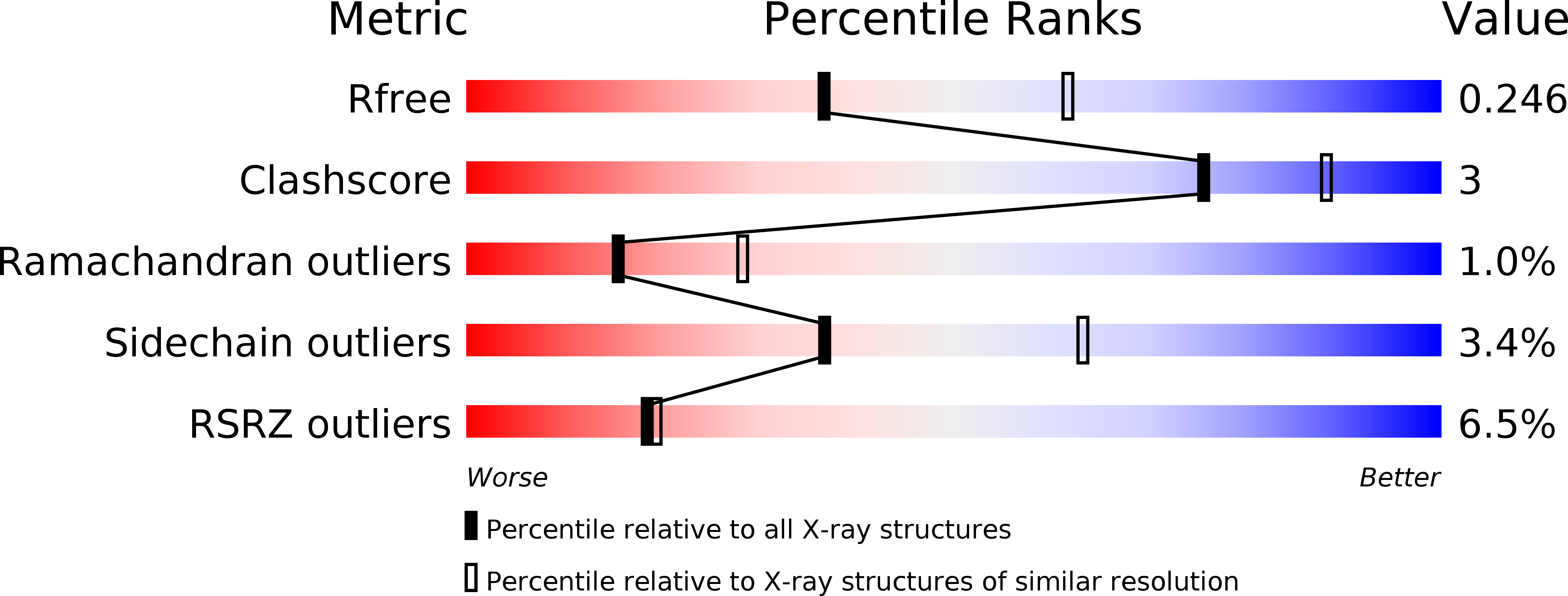
Resolution:
2.50 Å
R-Value Free:
0.25
R-Value Work:
0.21
R-Value Observed:
0.21
Space Group:
H 3
Macromolecular Entities
Polymer Type:polypeptide(L)
Molecule:CG5884-PA
Chain IDs:A
Chain Length:102
Number of Molecules:1
Biological Source:Drosophila melanogaster
Polymer Type:polypeptide(L)
Molecule:Pals1 peptide
Chain IDs:B
Chain Length:12
Number of Molecules:1
Biological Source:
Primary Citation
Internal recognition through PDZ domain plasticity in the Par-6-Pals1 complex.
Nat.Struct.Mol.Biol. 11 1122 1127 (2004)
PMID: 15475968 DOI: 10.1038/nsmb839

Abstact

PDZ protein interaction domains are typically selective for C-terminal ligands, but non-C-terminal, 'internal' ligands have also been identified. The PDZ domain from the cell polarity protein Par-6 binds C-terminal ligands and an internal sequence from the protein Pals1/Stardust. The structure of the Pals1-Par-6 PDZ complex reveals that the PDZ ligand-binding site is deformed to allow for internal binding. Whereas binding of the Rho GTPase Cdc42 to a CRIB domain adjacent to the Par-6 PDZ regulates binding of C-terminal ligands, the conformational change that occurs upon binding of Pals1 renders its binding independent of Cdc42. These results suggest a mechanism by which the requirement for a C terminus can be readily bypassed by PDZ ligands and reveal a complex set of cooperative and competitive interactions in Par-6 that are likely to be important for cell polarity regulation.

Legend

Protein

Chemical

Disease

Primary Citation of related structures