1X6V image
Entry Detail
PDB ID:
1X6V
Keywords:
Title:
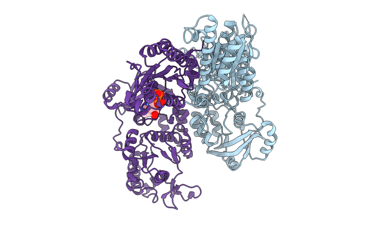
The crystal structure of human 3'-phosphoadenosine-5'-phosphosulfate synthetase 1
Biological Source:
Source Organism:
Host Organism:
PDB Version:
Deposition Date:
2004-08-12
Release Date:
2005-03-22
Method Details:
Experimental Method:
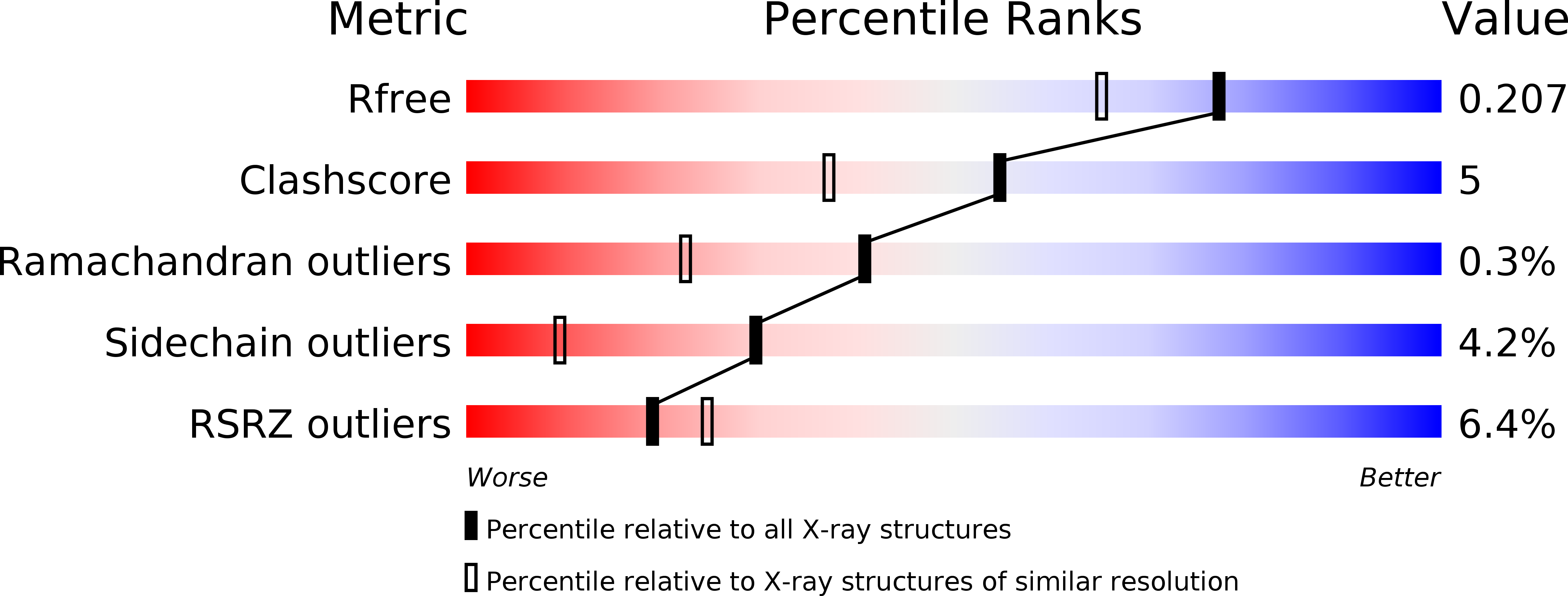
Resolution:
1.75 Å
R-Value Free:
0.19
R-Value Work:
0.16
R-Value Observed:
0.16
Space Group:
P 1 21 1
Macromolecular Entities
Polymer Type:polypeptide(L)
Description:Bifunctional 3'-phosphoadenosine 5'-phosphosulfate synthetase 1
Chain IDs:A (auth: B), B (auth: A)
Chain Length:630
Number of Molecules:2
Biological Source:Homo sapiens
Primary Citation
The Crystal Structure of Human PAPS Synthetase 1 Reveals Asymmetry in Substrate Binding
J.Mol.Biol. 347 623 635 (2005)
PMID: 15755455 DOI: 10.1016/j.jmb.2005.01.005

Abstact

The high energy sulfate donor 3'-phosphoadenosine-5-phosphosulfate (PAPS) is used for sulfate conjugation of extracellular matrix, hormones and drugs. Human PAPS synthetase 1 catalyzes two subsequent reactions starting from ATP and sulfate. First the ATP sulfurylase domain forms APS, then the APS kinase domain phosphorylates the APS intermediate to PAPS. Up to now the interaction between the two enzymatic activities remained elusive, mainly because of missing structural information. Here we present the crystal structure of human PAPSS1 at 1.8 angstroms resolution. The structure reveals a homodimeric, asymmetric complex with the shape of a chair. The two kinase domains adopt different conformational states, with only one being able to bind its two substrates. The asymmetric binding of ADP to the APS kinase is not only observed in the crystal structure, but can also be detected in solution, using an enzymatic assay. These observations strongly indicate structural changes during the reaction cycle. Furthermore crystals soaked with ADP and APS could be prepared and the corresponding structures could be solved.

Legend

Protein

Chemical

Disease

Primary Citation of related structures