1X3X image
Deposition Date 2005-05-11
Release Date 2006-06-06
Last Version Date 2024-10-30
Entry Detail
PDB ID:
1X3X
Title:
Crystal Structure of Cytochrome b5 from Ascaris suum
Biological Source:
Source Organism:
Ascaris suum (Taxon ID: 6253)
Method Details:
Experimental Method:
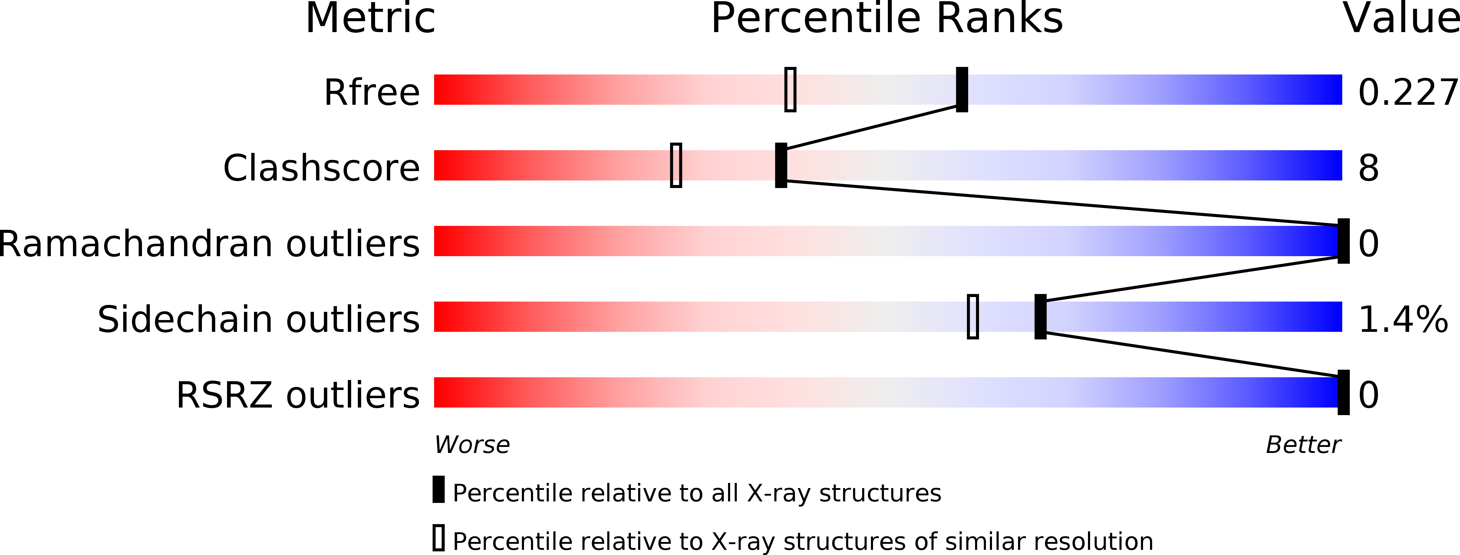
Resolution:
1.80 Å
R-Value Free:
0.23
R-Value Work:
0.19
Space Group:
C 1 2 1
Macromolecular Entities
Structures with similar UniProt ID
Protein Blast
Polymer Type:polypeptide(L)
Molecule:Cytochrome b5
Chain IDs:A, B
Chain Length:82
Number of Molecules:2
Biological Source:Ascaris suum
Primary Citation
Unique structure of Ascaris suum b5-type cytochrome: an additional alpha-helix and positively charged residues on the surface domain interact with redox partners
Biochem.J. 394 437 447 (2006)
PMID: 16288599 DOI: 10.1042/BJ20051308

Abstact

Cytochrome b5 of the body wall of adult Ascaris suum, a porcine parasitic nematode, is a soluble protein that lacks a C-terminal membrane-anchoring domain, but possesses an N-terminal pre-sequence of 30 amino acids. During the maturation of cytochrome b5, the N-terminal pre-sequence is proteolytically cleaved to form the mature protein of 82 amino acid residues. A. suum cytochrome b5 is a basic protein containing more lysine residues and exhibiting a higher midpoint redox potential than its mammalian counterparts. We developed an expression system for the production of the recombinant nematode cytochrome b5, which is chemically and functionally identical with the native protein. Using this recombinant protein, we have determined the X-ray crystal structure of A. suum cytochrome b5 at 1.8 A (1 A=0.1 nm) resolution, and we have shown that this protein is involved in the reduction of nematode body-wall metmyoglobin. The crystal structure of A. suum cytochrome b5 consists of six alpha-helices and five beta-strands. It differs from its mammalian counterparts by having a head-to-tail disulphide bridge, as well as a four-residue insertion in the vicinity of the sixth ligating histidine, which forms an additional alpha-helix, alpha4A, between helices alpha4 and alpha5. A. suum cytochrome b5 exists predominantly as a haem-orientation B isomer. Furthermore, the haem plane is rotated approx. 80 degrees relative to the axis formed by haem-Fe and N atoms of the two histidine residues that are ligated to haem-Fe. The charge distribution around the haem crevice of A. suum cytochrome b5 is remarkably different from that of mammalian cytochrome b5 in that the nematode protein bears positively charged lysine residues surrounding the haem crevice. Using immunohistochemistry, we found that A. suum cytochrome b5 is present in the nematode hypodermis. Based on this histochemical and structural information, the physiological function of A. suum cytochrome b5 and its interaction with nematode metmyoglobin can be hypothesized.

Legend

Protein

Chemical

Disease

Primary Citation of related structures