1X2H image
Deposition Date 2005-04-23
Release Date 2005-08-02
Last Version Date 2024-10-16
Entry Detail
PDB ID:
1X2H
Keywords:
Title:
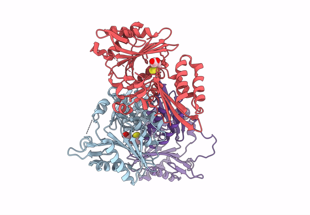
Crystal Structure of Lipate-Protein Ligase A from Escherichia coli complexed with lipoic acid
Biological Source:
Source Organism:
Escherichia coli (Taxon ID: 562)
Host Organism:
Method Details:
Experimental Method:
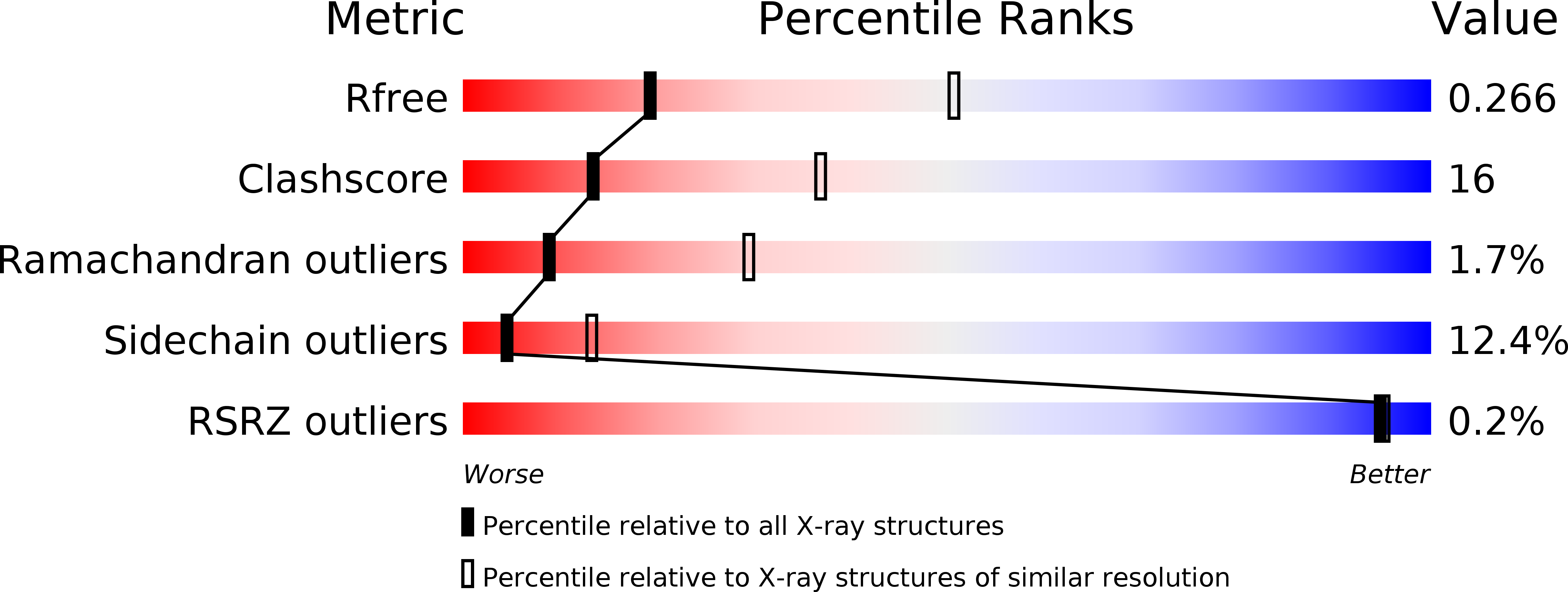
Resolution:
2.91 Å
R-Value Free:
0.27
R-Value Work:
0.18
R-Value Observed:
0.19
Space Group:
C 2 2 21
Macromolecular Entities
Polymer Type:polypeptide(L)
Molecule:Lipoate-protein ligase A
Gene (Uniprot):lplA
Chain IDs:A, B, C
Chain Length:337
Number of Molecules:3
Biological Source:Escherichia coli
Modified Residue
Compound ID Chain ID Parent Comp ID Details 2D Image
MSE A MET SELENOMETHIONINE
Ligand Molecules
Primary Citation
Crystal structure of lipoate-protein ligase A from Escherichia coli: Determination of the lipoic acid-binding site
J.Biol.Chem. 280 33645 33651 (2005)
PMID: 16043486 DOI: 10.1074/jbc.M505010200

Abstact

Lipoate-protein ligase A (LplA) catalyzes the formation of lipoyl-AMP from lipoate and ATP and then transfers the lipoyl moiety to a specific lysine residue on the acyltransferase subunit of alpha-ketoacid dehydrogenase complexes and on H-protein of the glycine cleavage system. The lypoyllysine arm plays a pivotal role in the complexes by shuttling the reaction intermediate and reducing equivalents between the active sites of the components of the complexes. We have determined the X-ray crystal structures of Escherichia coli LplA alone and in a complex with lipoic acid at 2.4 and 2.9 angstroms resolution, respectively. The structure of LplA consists of a large N-terminal domain and a small C-terminal domain. The structure identifies the substrate binding pocket at the interface between the two domains. Lipoic acid is bound in a hydrophobic cavity in the N-terminal domain through hydrophobic interactions and a weak hydrogen bond between carboxyl group of lipoic acid and the Ser-72 or Arg-140 residue of LplA. No large conformational change was observed in the main chain structure upon the binding of lipoic acid.

Legend

Protein

Chemical

Disease

Primary Citation of related structures