1X1V image
Deposition Date 2005-04-14
Release Date 2005-11-08
Last Version Date 2023-10-25
Entry Detail
PDB ID:
1X1V
Title:
Structure Of Banana Lectin- Methyl-Alpha-Mannose Complex
Biological Source:
Source Organism:
Musa acuminata (Taxon ID: 4641)
Method Details:
Experimental Method:
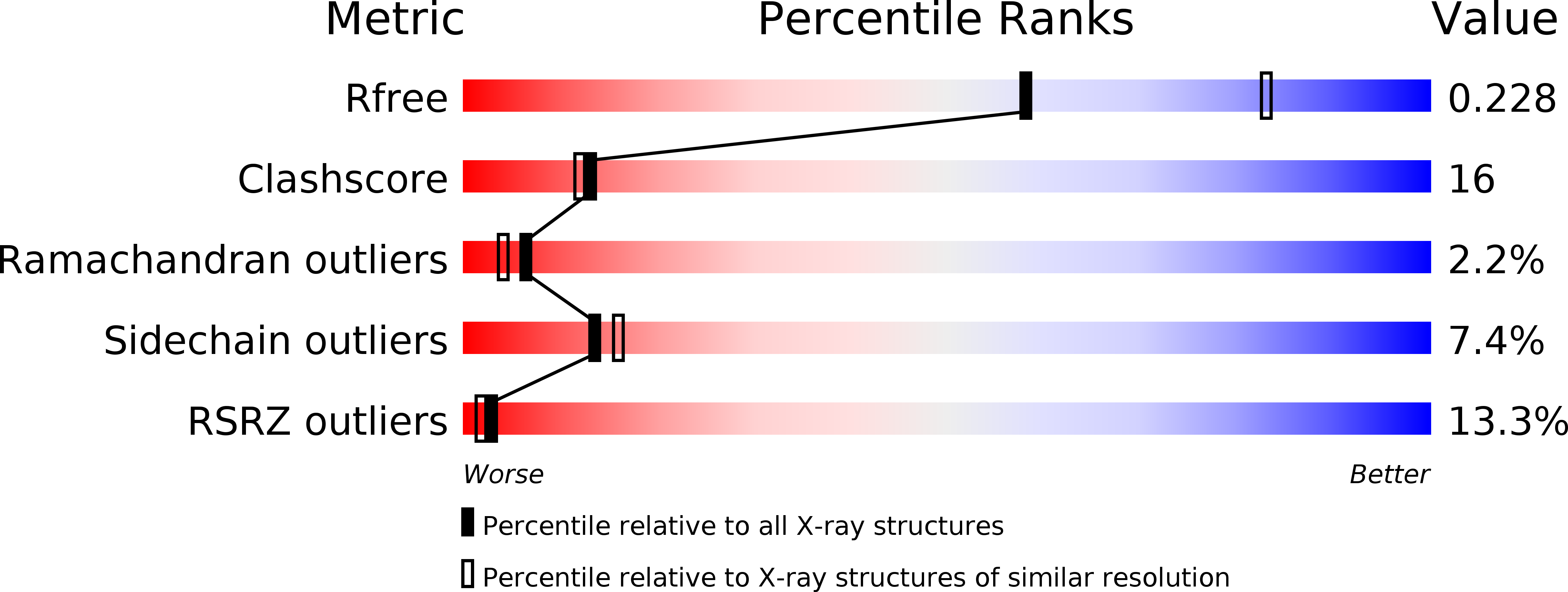
Resolution:
2.45 Å
R-Value Free:
0.26
R-Value Work:
0.22
R-Value Observed:
0.22
Space Group:
P 32 2 1
Macromolecular Entities
Structures with similar UniProt ID
Protein Blast
Polymer Type:polypeptide(L)
Molecule:lectin
Chain IDs:A, B
Chain Length:141
Number of Molecules:2
Biological Source:Musa acuminata
Primary Citation
Unusual sugar specificity of banana lectin from Musa paradisiaca and its probable evolutionary origin. Crystallographic and modelling studies
Glycobiology 15 1025 1032 (2005)
PMID: 15958419 DOI: 10.1093/glycob/cwi087

Abstact

The crystal structure of a complex of methyl-alpha-D-mannoside with banana lectin from Musa paradisiaca reveals two primary binding sites in the lectin, unlike in other lectins with beta-prism I fold which essentially consists of three Greek key motifs. It has been suggested that the fold evolved through successive gene duplication and fusion of an ancestral Greek key motif. In other lectins, all from dicots, the primary binding site exists on one of the three motifs in the three-fold symmetric molecule. Banana is a monocot, and the three motifs have not diverged enough to obliterate sequence similarity among them. Two Greek key motifs in it carry one primary binding site each. A common secondary binding site exists on the third Greek key. Modelling shows that both the primary sites can support 1-2, 1-3, and 1-6 linked mannosides with the second residue interacting in each case primarily with the secondary binding site. Modelling also readily leads to a bound branched mannopentose with the nonreducing ends of the two branches anchored at the two primary binding sites, providing a structural explanation for the lectin's specificity for branched alpha-mannans. A comparison of the dimeric banana lectin with other beta-prism I fold lectins, provides interesting insights into the variability in their quaternary structure.

Legend

Protein

Chemical

Disease

Primary Citation of related structures