1X1T image
Deposition Date 2005-04-13
Release Date 2006-01-10
Last Version Date 2023-10-25
Entry Detail
PDB ID:
1X1T
Keywords:
Title:
Crystal Structure of D-3-Hydroxybutyrate Dehydrogenase from Pseudomonas fragi Complexed with NAD+
Biological Source:
Source Organism:
Host Organism:
Method Details:
Experimental Method:
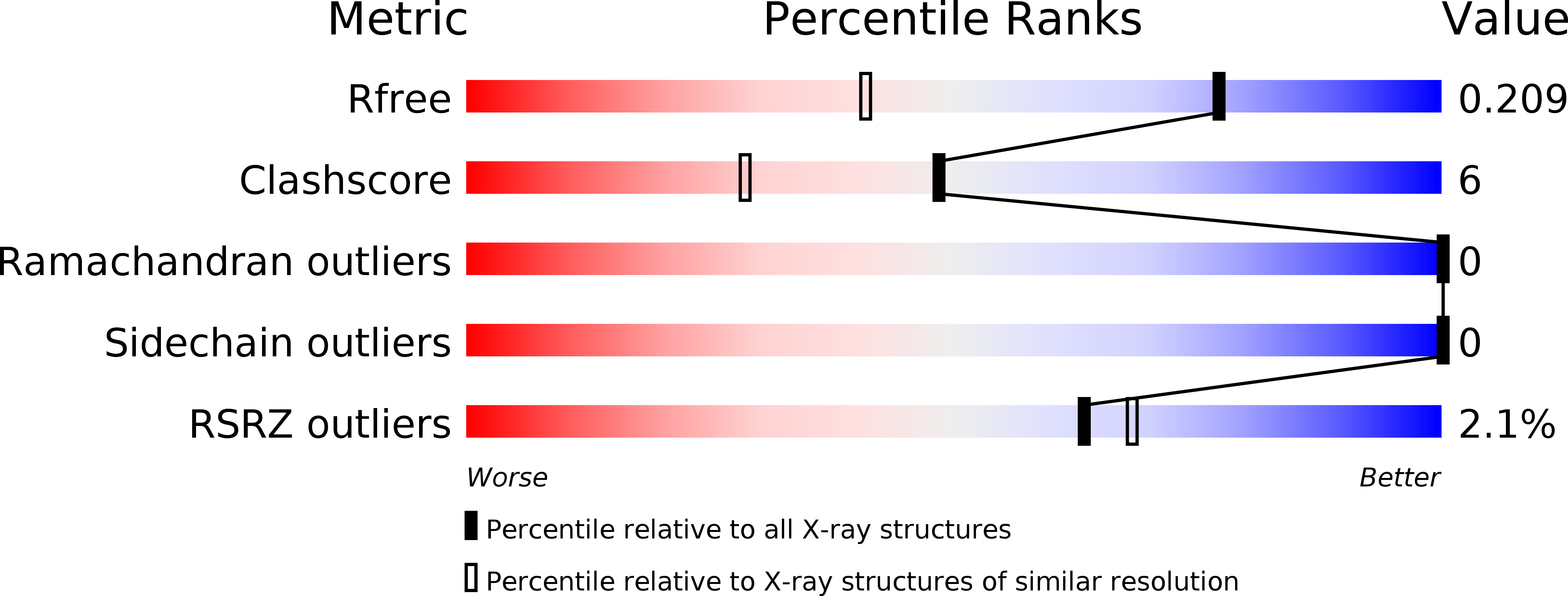
Resolution:
1.52 Å
R-Value Free:
0.21
R-Value Work:
0.19
Space Group:
I 2 2 2
Macromolecular Entities
Polymer Type:polypeptide(L)
Molecule:D(-)-3-hydroxybutyrate dehydrogenase
Chain IDs:A
Chain Length:260
Number of Molecules:1
Biological Source:Pseudomonas fragi
Primary Citation
d-3-Hydroxybutyrate Dehydrogenase from Pseudomonas fragi: Molecular Cloning of the Enzyme Gene and Crystal Structure of the Enzyme
J.Mol.Biol. 355 722 733 (2006)
PMID: 16325199 DOI: 10.1016/j.jmb.2005.10.072

Abstact

The gene coding for d-3-hydroxybutyrate dehydrogenase (HBDH) was cloned from Pseudomonas fragi. The nucleotide sequence contained a 780 bp open reading frame encoding a 260 amino acid residue protein. The recombinant enzyme was efficiently expressed in Escherichia coli cells harboring pHBDH11 and was purified to homogeneity as judged by SDS-PAGE. The enzyme showed a strict stereospecificity to the D-enantiomer (3R-configuration) of 3-hydroxybutyrate as a substrate. Crystals of the ligand-free HBDH and of the enzyme-NAD+ complex were obtained using the hanging-drop, vapor-diffusion method. The crystal structure of the HBDH was solved by the multiwavelength anomalous diffraction method using the SeMet-substituted enzyme and was refined to 2.0 A resolution. The overall structure of P.fragi HBDH, including the catalytic tetrad of Asn114, Ser142, Tyr155, and Lys159, shows obvious relationships with other members of the short-chain dehydrogenase/reductase (SDR) family. A cacodylate anion was observed in both the ligand-free enzyme and the enzyme-NAD+ complex, and was located near the catalytic tetrad. It was shown that the cacodylate inhibited the NAD+-dependent D-3-hydroxybutyrate dehydrogenation competitively, with a Ki value of 5.6 mM. From the interactions between cacodylate and the enzyme, it is predicted that substrate specificity is achieved through the recognition of the 3-methyl and carboxyl groups of the substrate.

Legend

Protein

Chemical

Disease

Primary Citation of related structures