Deposition Date
2005-03-28
Release Date
2005-08-30
Last Version Date
2024-10-23
Entry Detail
PDB ID:
1X0S
Keywords:
Title:
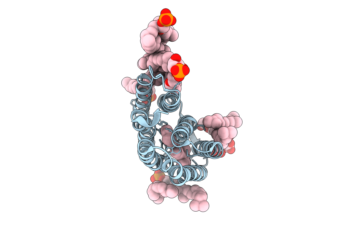
Crystal structure of the 13-cis isomer of bacteriorhodopsin
Biological Source:
Source Organism(s):
Halobacterium salinarum (Taxon ID: 2242)
Method Details:
Experimental Method:
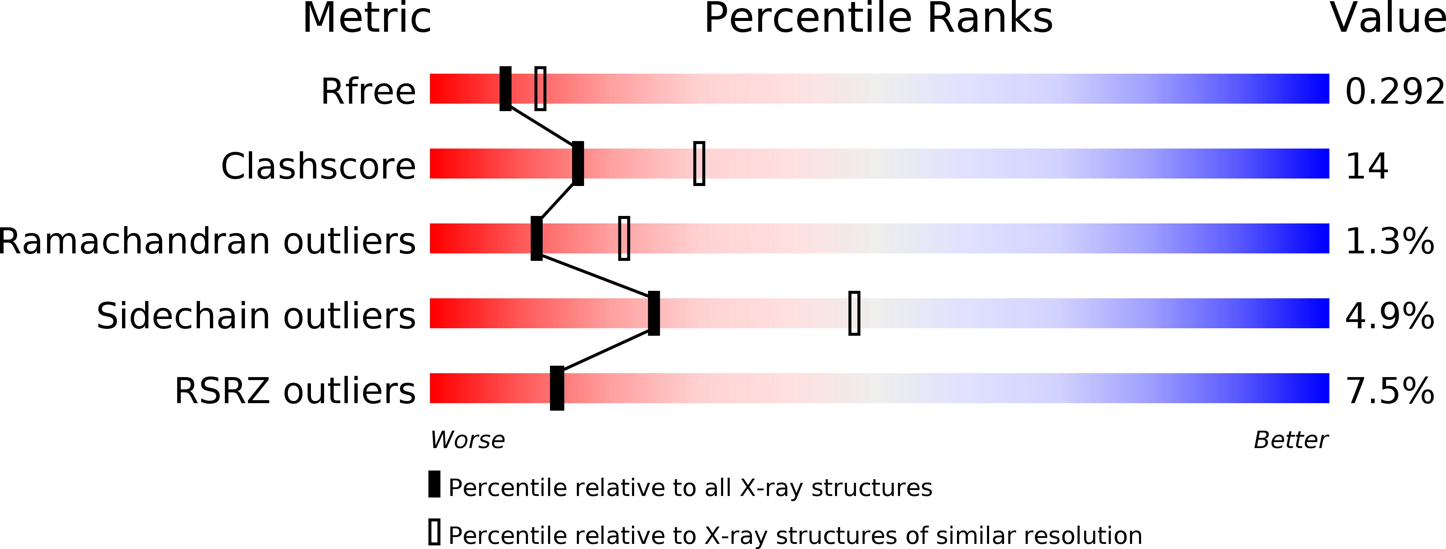
Resolution:
2.50 Å
R-Value Free:
0.30
R-Value Work:
0.27
R-Value Observed:
0.27
Space Group:
P 6 2 2