1X0P image
Deposition Date 2005-03-27
Release Date 2005-06-07
Last Version Date 2024-03-13
Entry Detail
PDB ID:
1X0P
Title:
Structure of a cyanobacterial BLUF protein, Tll0078
Biological Source:
Source Organism(s):
Expression System(s):
Method Details:
Experimental Method:
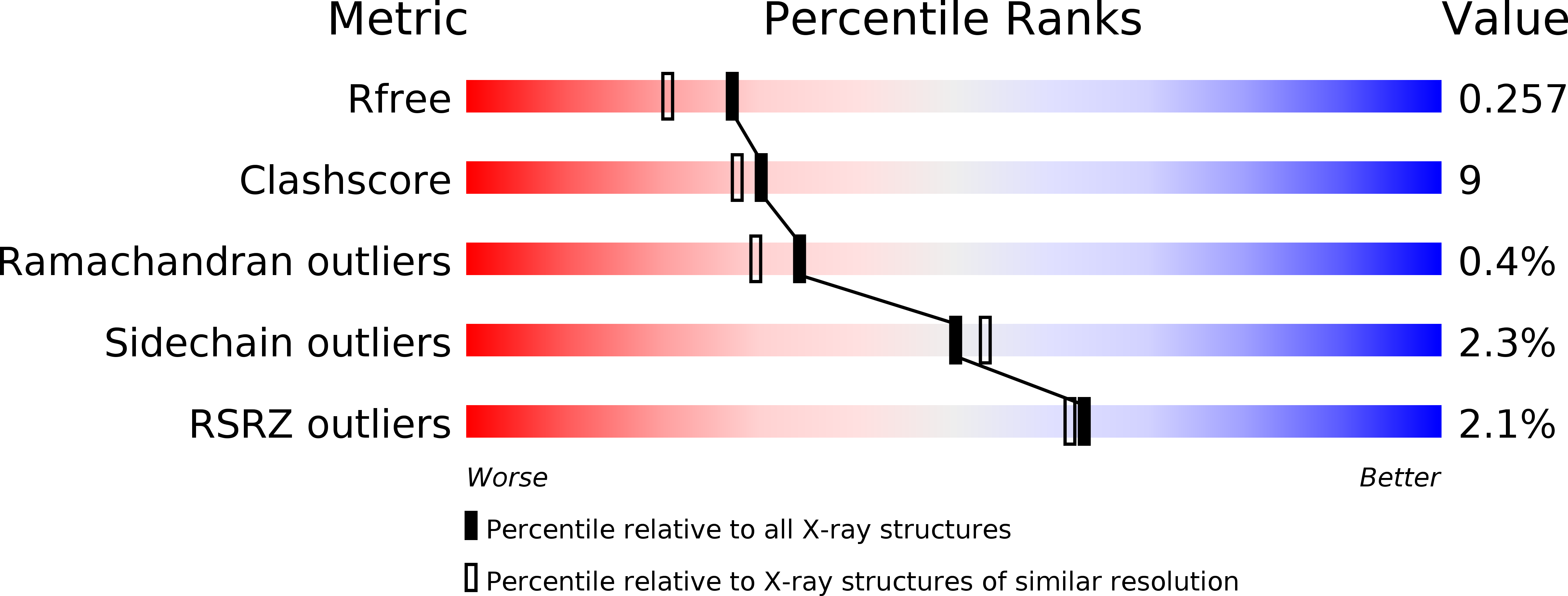
Resolution:
2.00 Å
R-Value Free:
0.26
R-Value Work:
0.23
Space Group:
P 21 21 21
Macromolecular Entities
Structures with similar UniProt ID
Protein Blast
Polymer Type:polypeptide(L)
Molecule:hypothetical protein Tll0078
Gene (Uniprot):tll0078
Chain IDs:A, B, C, D, E, F, G, H, I, J
Chain Length:143
Number of Molecules:10
Biological Source:Thermosynechococcus elongatus
Ligand Molecules
Primary Citation
Structure of a Cyanobacterial BLUF Protein, Tll0078, Containing a Novel FAD-binding Blue Light Sensor Domain
J.Mol.Biol. 349 1 9 (2005)
PMID: 15876364 DOI: 10.1016/j.jmb.2005.03.067

Abstact

The sensor proteins for blue light using the FAD (BLUF) domain belong to the third family of the photoreceptor proteins using a flavin chromophore, where the other two families are phototropins and cryptochromes. As the first structure of this BLUF domain, we have determined the crystal structure of the Tll0078 protein from Thermosynechococcus elongatus BP-1, which contains a BLUF domain bound to FAD, at 2A resolution. Five Tll0078 monomers are located around the non-crystallographic 5-fold axis to form a pentamer, and two pentamers related by 2-fold non-crystallographic symmetry form a decameric assembly. The monomer consists of two domains, the BLUF domain at the N-terminal region and the C-terminal domain. The overall structure of the BLUF domain consists of a five-stranded mixed beta-sheet with two alpha-helices running parallel with it. The isoalloxazine ring of FAD is accommodated in a pocket formed by several highly conserved amino acid residues in the BLUF domain. Of these, the three apparent key residues (Asn31, Asn32 and Gln50) were substituted with Ala. Mutant proteins of N31A and N32A showed a nearly normal 10nm spectral shift of the flavin upon illumination, while the Q50A mutant did not exhibit such a shift at all. On the basis of the crystal structure, we discussed a possible role of Gln50, which is structurally and functionally linked with the critical Tyr8 (FAD-Gln50-Tyr8 network), with regard to the light-induced spectral shift of the BLUF proteins.

Legend

Protein

Chemical

Disease

Primary Citation of related structures
Feedback Form
Name
Email
Institute
Feedback