1X0L image
Deposition Date 2005-03-24
Release Date 2005-10-04
Last Version Date 2024-03-13
Entry Detail
PDB ID:
1X0L
Keywords:
Title:
Crystal structure of tetrameric homoisocitrate dehydrogenase from an extreme thermophile, Thermus thermophilus
Biological Source:
Source Organism(s):
Expression System(s):
Method Details:
Experimental Method:
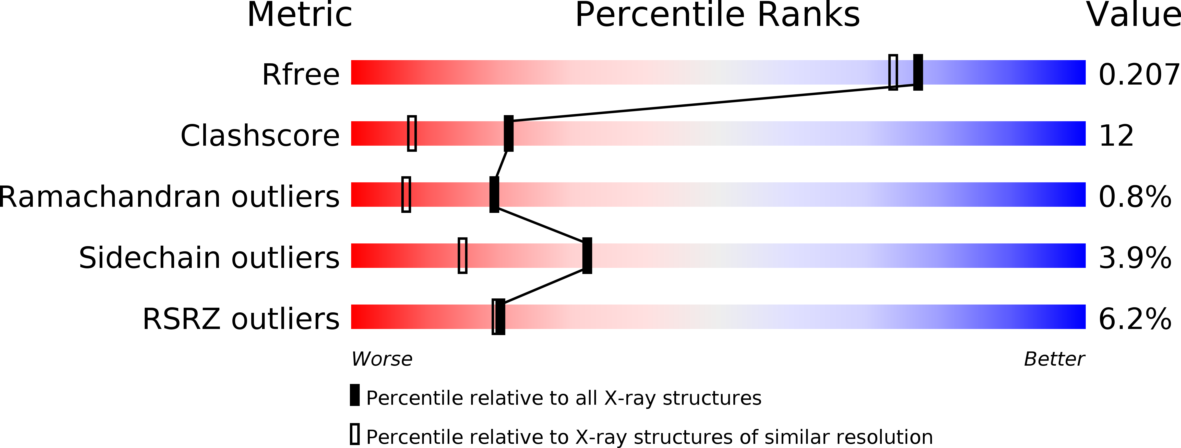
Resolution:
1.85 Å
R-Value Free:
0.24
R-Value Work:
0.21
Space Group:
C 2 2 21
Macromolecular Entities
Structures with similar UniProt ID
Protein Blast
Polymer Type:polypeptide(L)
Molecule:Homoisocitrate dehydrogenase
Chain IDs:A, B
Chain Length:333
Number of Molecules:2
Biological Source:Thermus thermophilus
Primary Citation
Crystal Structure of Tetrameric Homoisocitrate Dehydrogenase from an Extreme Thermophile, Thermus thermophilus: Involvement of Hydrophobic Dimer-Dimer Interaction in Extremely High Thermotolerance
J.Bacteriol. 187 6779 6788 (2005)
PMID: 16166541 DOI: 10.1128/JB.187.19.6779-6788.2005

Abstact

The crystal structure of homoisocitrate dehydrogenase involved in lysine biosynthesis from Thermus thermophilus (TtHICDH) was determined at 1.85-A resolution. Arg85, which was shown to be a determinant for substrate specificity in our previous study, is positioned close to the putative substrate binding site and interacts with Glu122. Glu122 is highly conserved in the equivalent position in the primary sequence of ICDH and archaeal 3-isopropylmalate dehydrogenase (IPMDH) but interacts with main- and side-chain atoms in the same domain in those paralogs. In addition, a conserved Tyr residue (Tyr125 in TtHICDH) which extends its side chain toward a substrate and thus has a catalytic function in the related beta-decarboxylating dehydrogenases, is flipped out of the substrate-binding site. These results suggest the possibility that the conformation of the region containing Glu122-Tyr125 is changed upon substrate binding in TtHICDH. The crystal structure of TtHICDH also reveals that the arm region is involved in tetramer formation via hydrophobic interactions and might be responsible for the high thermotolerance. Mutation of Val135, located in the dimer-dimer interface and involved in the hydrophobic interaction, to Met alters the enzyme to a dimer (probably due to steric perturbation) and markedly decreases the thermal inactivation temperature. Both the crystal structure and the mutation analysis indicate that tetramer formation is involved in the extremely high thermotolerance of TtHICDH.

Legend

Protein

Chemical

Disease

Primary Citation of related structures
Feedback Form
Name
Email
Institute
Feedback