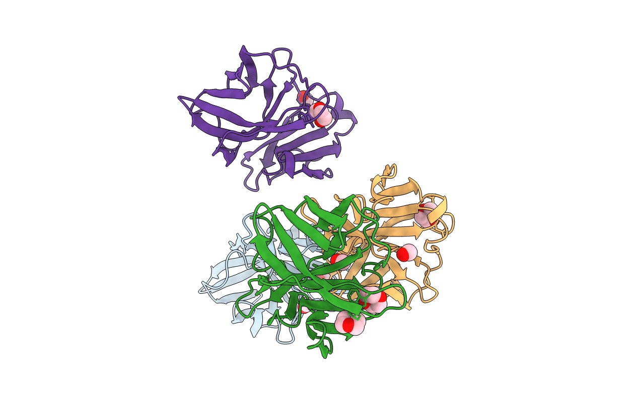
1WYK image
Entry Detail
PDB ID:
1WYK
Title:
SINDBIS VIRUS CAPSID PROTEIN (114-264)
Biological Source:
Source Organism:
PDB Version:
Deposition Date:
1998-01-12
Release Date:
1998-04-29
Method Details:
Experimental Method:
Resolution:
2.00 Å
R-Value Free:
0.26
R-Value Work:
0.19
R-Value Observed:
0.19
Space Group:
P 1
Macromolecular Entities
Polymer Type:polypeptide(L)
Description:SINDBIS VIRUS CAPSID PROTEIN
Chain IDs:A, B, C, D
Chain Length:152
Number of Molecules:4
Biological Source:Sindbis virus
Primary Citation
Probing the potential glycoprotein binding site of sindbis virus capsid protein with dioxane and model building.
Proteins 33 311 317 (1998)
PMID: 9779796 DOI: 10.1002/(SICI)1097-0134(19981101)33:2<311::AID-PROT13>3.3.CO;2-L

Abstact

Alphavirus budding from the plasma membrane is initiated by the specific interaction of the nucleocapsid with the cytoplasmic domain of the glycoprotein E2. It was proposed (Lee et al., Structure 4:531-541, 1996) that binding of the capsid protein residues 108 to 110 (the "N-terminal arm" residues) to a hydrophobic pocket on the surface of the neighboring capsid protein in the crystal structure mimics the binding of the E2 C-terminal residues into this pocket. In addition, structural comparisons of wild-type and mutant Sindbis virus capsid protein (SCP) and Semliki Forest virus capsid protein suggested that budding is associated with a switch between two conformations of the hydrophobic pocket. To test the proposed mechanism, SCP(114-264), which is missing the N-terminal arm, was crystallized to examine the pocket conformation when the pocket is empty. However, the pocket was occupied by dioxane molecules from the crystallization solution. The pocket conformation was the same as that when it was occupied by the N-terminal arm, demonstrating that the pocket favors binding ligands of appropriate size and shape.

Legend

Protein

Chemical

Disease

Primary Citation of related structures