1WXR image
Deposition Date 2005-01-31
Release Date 2005-03-01
Last Version Date 2024-03-13
Entry Detail
PDB ID:
1WXR
Keywords:
Title:
Crystal structure of Heme Binding protein, an autotransporter hemoglobine protease from pathogenic Escherichia coli
Biological Source:
Source Organism:
Escherichia coli (Taxon ID: 562)
Host Organism:
Method Details:
Experimental Method:
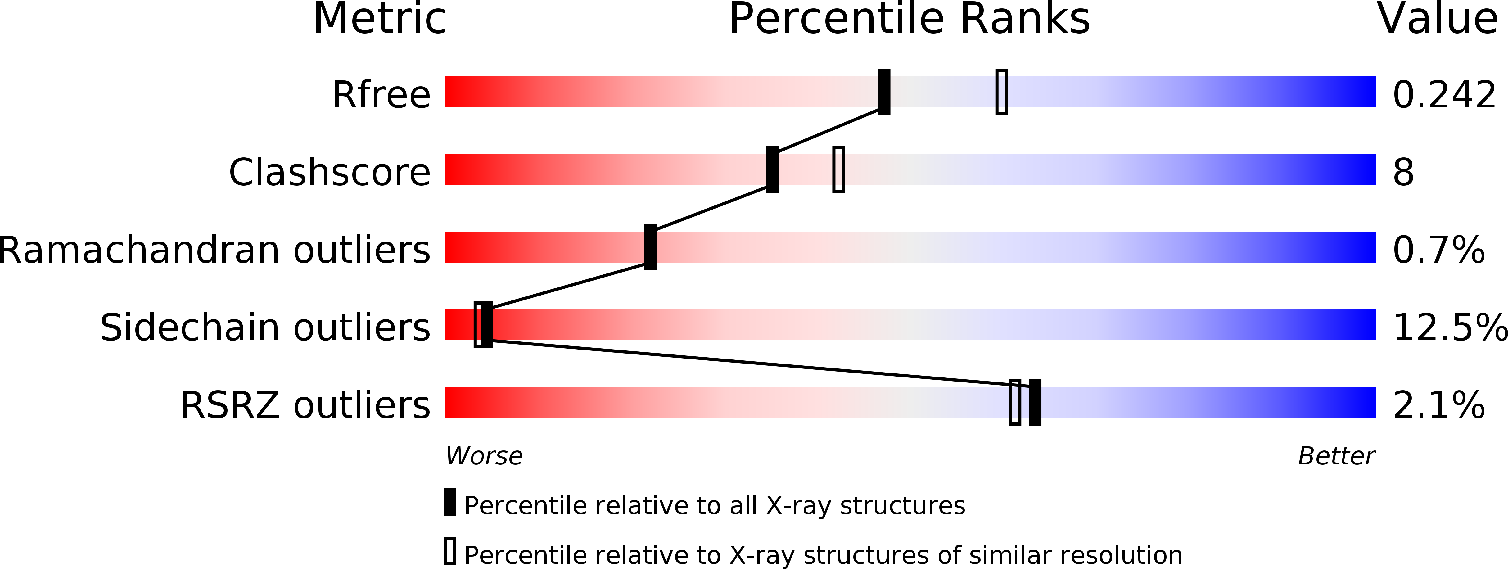
Resolution:
2.20 Å
R-Value Free:
0.24
R-Value Work:
0.20
R-Value Observed:
0.20
Space Group:
P 61 2 2
Macromolecular Entities
Structures with similar UniProt ID
Protein Blast
Polymer Type:polypeptide(L)
Molecule:haemoglobin protease
Gene (Uniprot):hbp
Chain IDs:A
Chain Length:1048
Number of Molecules:1
Biological Source:Escherichia coli
Primary Citation
Crystal structure of heme binding protein, an autotransporter hemoglobin protease from pathogenic escherichia coli
J.Biol.Chem. 280 17339 17345 (2005)
PMID: 15728184 DOI: 10.1074/jbc.M412885200

Abstact

The acquisition of iron is essential for the survival of pathogenic bacteria, which have consequently evolved a wide variety of uptake systems to extract iron and heme from host proteins such as hemoglobin. Hemoglobin protease (Hbp) was discovered as a factor involved in the symbiosis of pathogenic Escherichia coli and Bacteroides fragilis, which cause intra-abdominal abscesses. Released from E. coli, this serine protease autotransporter degrades hemoglobin and delivers heme to both bacterial species. The crystal structure of the complete passenger domain of Hbp (110 kDa) is presented, which is the first structure from this class of serine proteases and the largest parallel beta-helical structure yet solved.

Legend

Protein

Chemical

Disease

Primary Citation of related structures