1WVQ image
Entry Detail
PDB ID:
1WVQ
Title:
Structure of conserved hypothetical protein PAE2307 from Pyrobaculum aerophilum
Biological Source:
Source Organism:
PDB Version:
Deposition Date:
2004-12-24
Release Date:
2006-01-10
Method Details:
Experimental Method:
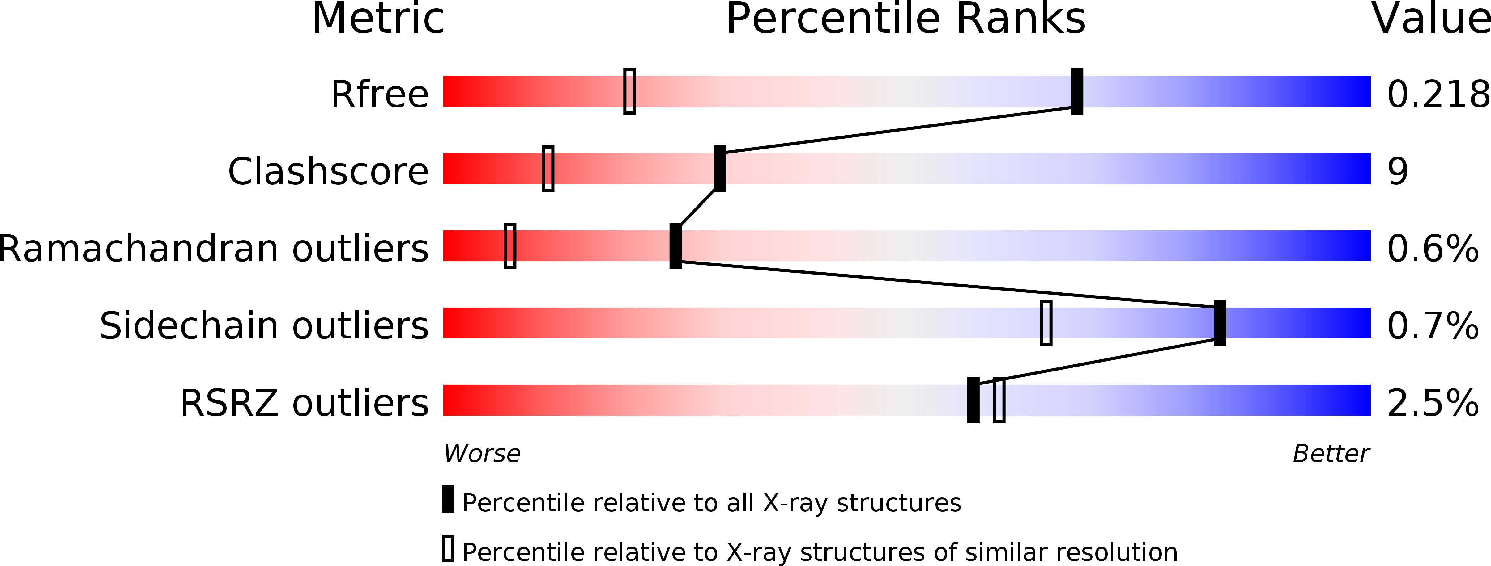
Resolution:
1.45 Å
R-Value Free:
0.22
R-Value Work:
0.20
R-Value Observed:
0.20
Space Group:
I 41 2 2
Macromolecular Entities
Polymer Type:polypeptide(L)
Description:hypothetical protein PAE2307
Chain IDs:A, B, C
Chain Length:167
Number of Molecules:3
Biological Source:Pyrobaculum aerophilum
Modified Residue
Compound ID Chain ID Parent Comp ID Details 2D Image
NEP A HIS N1-PHOSPHONOHISTIDINE
Ligand Molecules
Primary Citation
The structure of an ancient conserved domain establishes a structural basis for stable histidine phosphorylation and identifies a new family of adenosine-specific kinases.
J.Biol.Chem. 281 22131 22141 (2006)
PMID: 16737961 DOI: 10.1074/jbc.M603062200

Abstact

Phosphorylation of both small molecules and proteins plays a central role in many biological processes. In proteins, phosphorylation most commonly targets the oxygen atoms of Ser, Thr, and Tyr. In contrast, stably phosphorylated His residues are rarely found, due to the lability of the N-P bond, and histidine phosphorylation features most often in transient processes. Here we present the crystal structure of a protein of previously unknown function, which proves to contain a stably phosphorylated histidine residue. The protein is the product of open reading frame PAE2307, from the hyperthermophilic archaeon Pyrobaculum aerophilum, and is representative of a highly conserved protein family found in archaea and bacteria. The crystal structure of PAE2307, solved at 1.45-A resolution (R = 0.208, R(free) = 0.227), forms a remarkably tightly associated hexamer. The phosphorylated histidine at the proposed active site, pHis85, occupies a cavity that is at the interface between two subunits and contains a number of fully conserved residues. Stable phosphorylation is attributed to favorable hydrogen bonding of the phosphoryl group and a salt bridge with pHis85 that provides electronic stabilization. In silico modeling suggested that the protein may function as an adenosine kinase, a conclusion that is supported by in vitro assays of adenosine binding, using fluorescence spectroscopy, and crystallographic visualization of an adenosine complex of PAE2307 at 2.25-A resolution.

Legend

Protein

Chemical

Disease

Primary Citation of related structures