1WUU image
Deposition Date 2004-12-08
Release Date 2004-12-28
Last Version Date 2024-10-23
Entry Detail
PDB ID:
1WUU
Keywords:
Title:
crystal structure of human galactokinase complexed with MgAMPPNP and galactose
Biological Source:
Source Organism:
Homo sapiens (Taxon ID: 9606)
Host Organism:
Method Details:
Experimental Method:
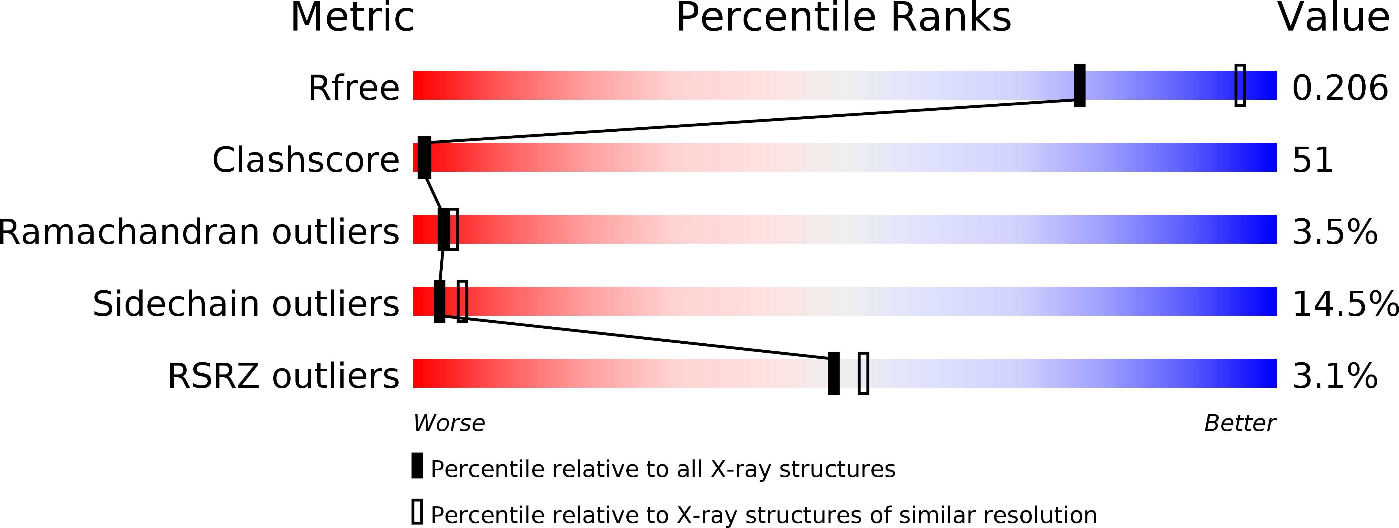
Resolution:
2.50 Å
R-Value Free:
0.25
R-Value Work:
0.20
R-Value Observed:
0.20
Space Group:
P 1 21 1
Macromolecular Entities
Structures with similar UniProt ID
Protein Blast
Polymer Type:polypeptide(L)
Molecule:Galactokinase
Gene (Uniprot):GALK1
Chain IDs:A, B, C, D
Chain Length:399
Number of Molecules:4
Biological Source:Homo sapiens
Modified Residue
Compound ID Chain ID Parent Comp ID Details 2D Image
MSE A MET SELENOMETHIONINE
Primary Citation
Molecular Structure of Human Galactokinase: IMPLICATIONS FOR TYPE II GALACTOSEMIA
J.Biol.Chem. 280 9662 9670 (2005)
PMID: 15590630 DOI: 10.1074/jbc.M412916200

Abstact

Galactokinase functions in the Leloir pathway for galactose metabolism by catalyzing the MgATP-dependent phosphorylation of the C-1 hydroxyl group of alpha-D-galactose. The enzyme is known to belong to the GHMP superfamily of small molecule kinases and has attracted significant research attention for well over 40 years. Approximately 20 mutations have now been identified in human galactokinase, which result in the diseased state referred to as Type II galactosemia. Here we report the three-dimensional architecture of human galactokinase with bound alpha-D-galactose and Mg-AMPPNP. The overall fold of the molecule can be described in terms of two domains with the active site wedged between them. The N-terminal domain is dominated by a six-stranded mixed beta-sheet whereas the C-terminal motif contains six alpha-helices and two layers of anti-parallel beta-sheet. Those residues specifically involved in sugar binding include Arg37, Glu43, His44, Asp46, Gly183, Asp186, and Tyr236. The C-1 hydroxyl group of alpha-D-galactose sits within 3.3 A of the gamma-phosphorus of the nucleotide and 3.4 A of the guanidinium group of Arg37. The carboxylate side chain of Asp186 lies within approximately 3.2 A of the C-2 hydroxyl group of alpha-D-galactose and the guanidinium group of Arg37. Both Arg37 and Asp186 are strictly conserved among both prokaryotic and eukaryotic galactokinases. In addition to providing molecular insight into the active site geometry of the enzyme, the model also provides a structural framework upon which to more fully understand the consequences of the those mutations known to give rise to Type II galactosemia.

Legend

Protein

Chemical

Disease

Primary Citation of related structures