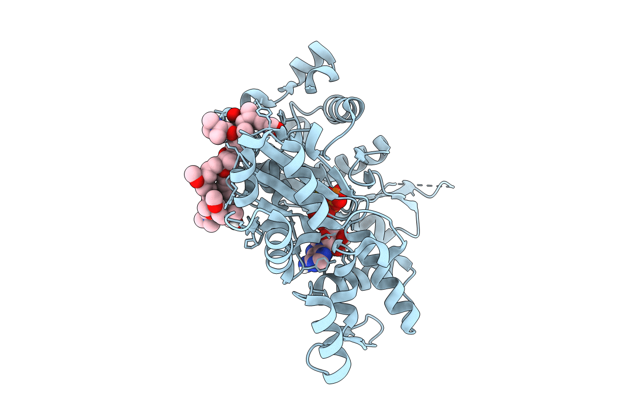
1WUA image
Deposition Date 2004-12-03
Release Date 2006-02-14
Last Version Date 2023-10-25
Entry Detail
PDB ID:
1WUA
Title:
The structure of Aplyronine A-actin complex
Biological Source:
Source Organism:
Method Details:
Experimental Method:
Resolution:
1.45 Å
R-Value Free:
0.17
R-Value Work:
0.15
R-Value Observed:
0.15
Space Group:
P 1 21 1
Macromolecular Entities
Polymer Type:polypeptide(L)
Molecule:Actin, alpha skeletal muscle
Gene (Uniprot):ACTA1
Chain IDs:A
Chain Length:375
Number of Molecules:1
Biological Source:Oryctolagus cuniculus
Modified Residue
Compound ID Chain ID Parent Comp ID Details 2D Image
HIC A HIS 4-METHYL-HISTIDINE
Primary Citation
Structure basis for antitumor effect of aplyronine a
J.Mol.Biol. 356 945 954 (2006)
PMID: 16406066 DOI: 10.1016/j.jmb.2005.12.031

Abstact

Aplyronine A, isolated from the sea hare Aplysia kurodai, possesses an exceedingly potent antitumor effect in vivo and it is one of the promising candidates as an anticancer drug. This macrolide is known to depolymerize F-actin and inhibit the polymerization of actin by forming a 1:1 complex with monomeric actin. The first complex structure of actin-aplyronine A was determined via a synchrotron X-ray analysis at a 1.45 A resolution. As expected, aplyronine A binds to a hydrophobic cleft composed of subdomains 1 and 3 of actin by intercalating its aliphatic tail part into the actin molecule as do the other reported F-actin depolymerizing agents. Unexpectedly, this complex structure shows the specific structural features around the trimethylserine moiety, revealed as an important moiety of aplyronine A for cytotoxicity against HeLa cells. Combining this result and our previous one, the moiety should strongly relate to the specific biological activity of aplyronine A; i.e. a potent antitumor effect.

Legend

Protein

Chemical

Disease

Primary Citation of related structures