Deposition Date
2004-11-23
Release Date
2005-03-08
Last Version Date
2023-10-25
Entry Detail
PDB ID:
1WTH
Keywords:
Title:
Crystal structure of gp5-S351L mutant and gp27 complex
Biological Source:
Source Organism(s):
Enterobacteria phage T4 (Taxon ID: 10665)
Expression System(s):
Method Details:
Experimental Method:
Resolution:
2.80 Å
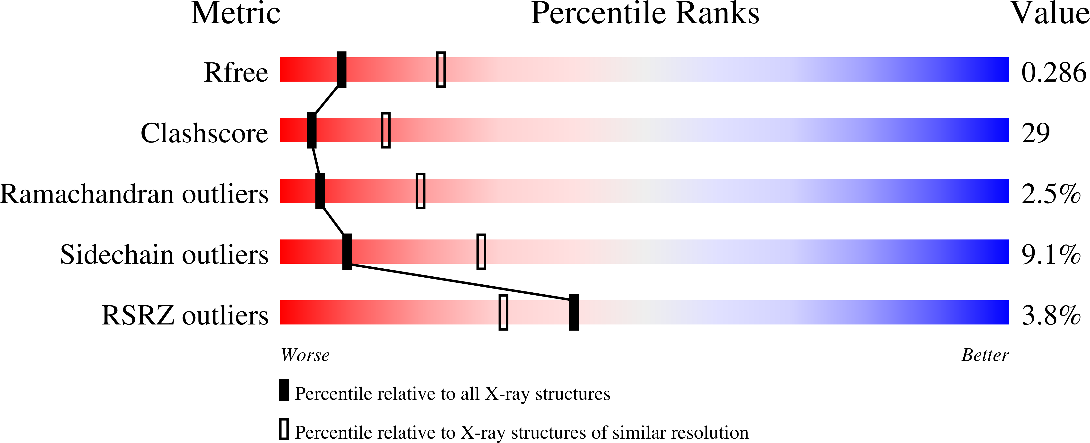
R-Value Free:
0.28
R-Value Work:
0.21
R-Value Observed:
0.21
Space Group:
H 3 2