1WS8 image
Deposition Date 2004-11-02
Release Date 2004-11-23
Last Version Date 2024-11-13
Entry Detail
PDB ID:
1WS8
Title:
Crystal Structure of Mavicyanin from Cucurbita pepo medullosa (Zucchini)
Biological Source:
Source Organism:
Cucurbita pepo (Taxon ID: 3663)
Host Organism:
Method Details:
Experimental Method:
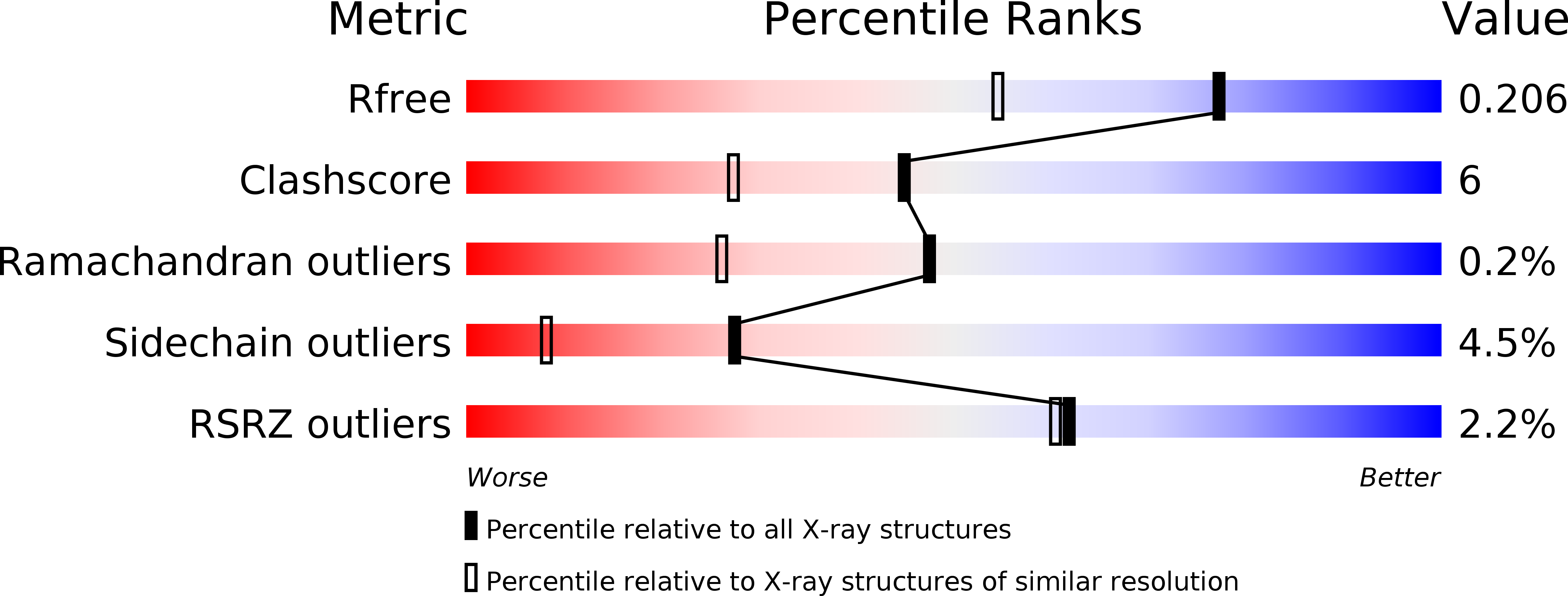
Resolution:
1.60 Å
R-Value Free:
0.21
R-Value Work:
0.19
R-Value Observed:
0.19
Space Group:
P 61
Macromolecular Entities
Polymer Type:polypeptide(L)
Molecule:mavicyanin
Chain IDs:A, B, C, D
Chain Length:109
Number of Molecules:4
Biological Source:Cucurbita pepo
Primary Citation
Structural reorganization of the copper binding site involving Thr15 of mavicyanin from Cucurbita pepo medullosa (zucchini) upon reduction.
J.Biochem.(Tokyo) 137 455 461 (2005)
PMID: 15858169 DOI: 10.1093/jb/mvi062

Abstact

Mavicyanin, a glycosylated protein isolated from Cucurbita pepo medullosa (zucchini), is a member of the phytocyanin subfamily containing one polypeptide chain of 109 amino residues and an unusual type-I Cu site in which the copper ligands are His45, Cys86, His91, and Gln96. The crystal structures of oxidized and reduced mavicyanin were determined at 1.6 and 1.9 A resolution, respectively. Mavicyanin has a core structure of seven polypeptide beta-strands arranged as a beta-sandwich organized into two beta-sheets, and the structure considerably resembles that of stellacyanin from cucumber (CST) or cucumber basic protein (CBP). A flexible region was not observed on superimpositioning of the oxidized and reduced mavicyanin structures. However, the Cu(II)-epsilon-O-Gln96 bond length was extended by 0.47 A, and the Thr15 residue was rotated by 60.0 degrees and O-gamma1-Thr15 moved from a distance of 4.78 to 2.58 A from the ligand Gln96 forming a new hydrogen bond between O-gamma1-Thr15 and epsilon-O-Gln96 upon reduction. The reorganization of copper coordination geometry of mavicyanin upon reduction arouses reduction potential decreased above pH 8 [Battistuzzi et al. (2001) J. Inorg. Biochem. 83, 223-227]. The rotation of Thr15 and the hydrogen bonding with the ligand Gln96 may constitute structural evidence of the decrease in the reduction potential at high pH.

Legend

Protein

Chemical

Disease

Primary Citation of related structures