Deposition Date
2004-10-12
Release Date
2005-06-28
Last Version Date
2024-10-16
Entry Detail
PDB ID:
1WR6
Keywords:
Title:
Crystal structure of GGA3 GAT domain in complex with ubiquitin
Biological Source:
Source Organism(s):
Homo sapiens (Taxon ID: 9606)
Bos taurus (Taxon ID: 9913)
Bos taurus (Taxon ID: 9913)
Expression System(s):
Method Details:
Experimental Method:
Resolution:
2.60 Å
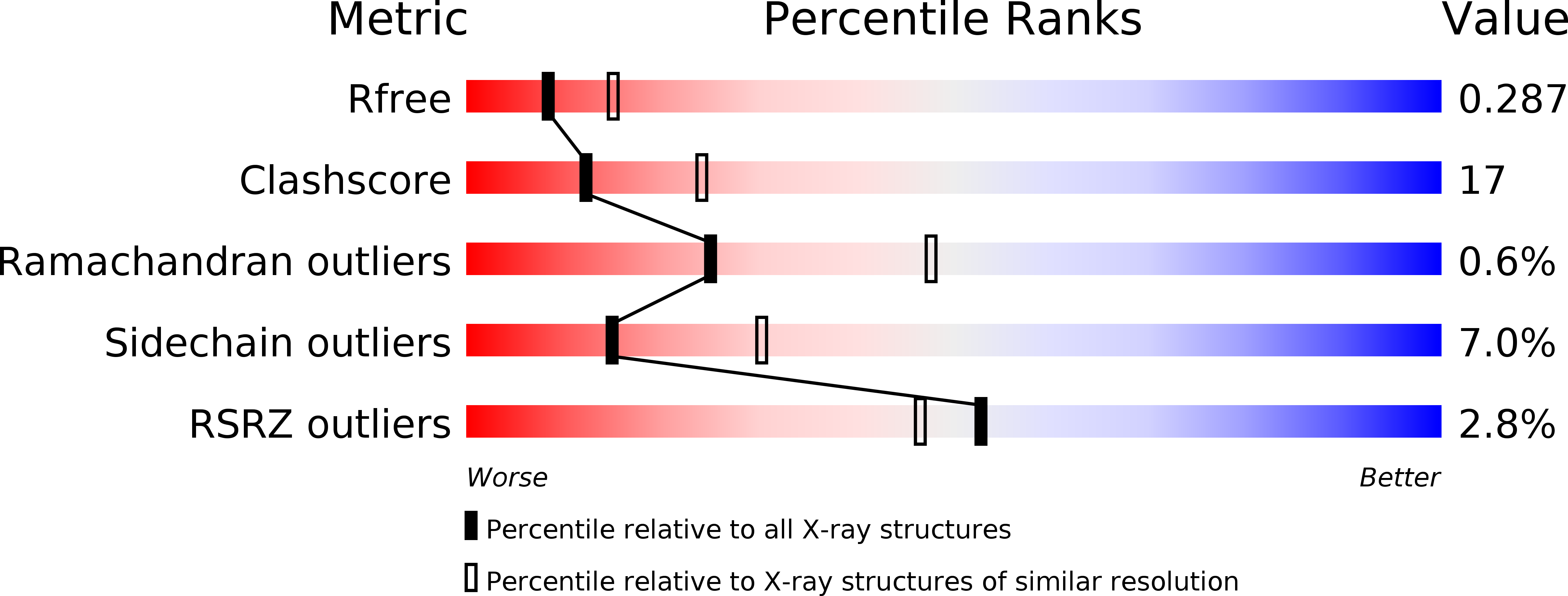
R-Value Free:
0.29
R-Value Work:
0.22
R-Value Observed:
0.22
Space Group:
P 21 21 21