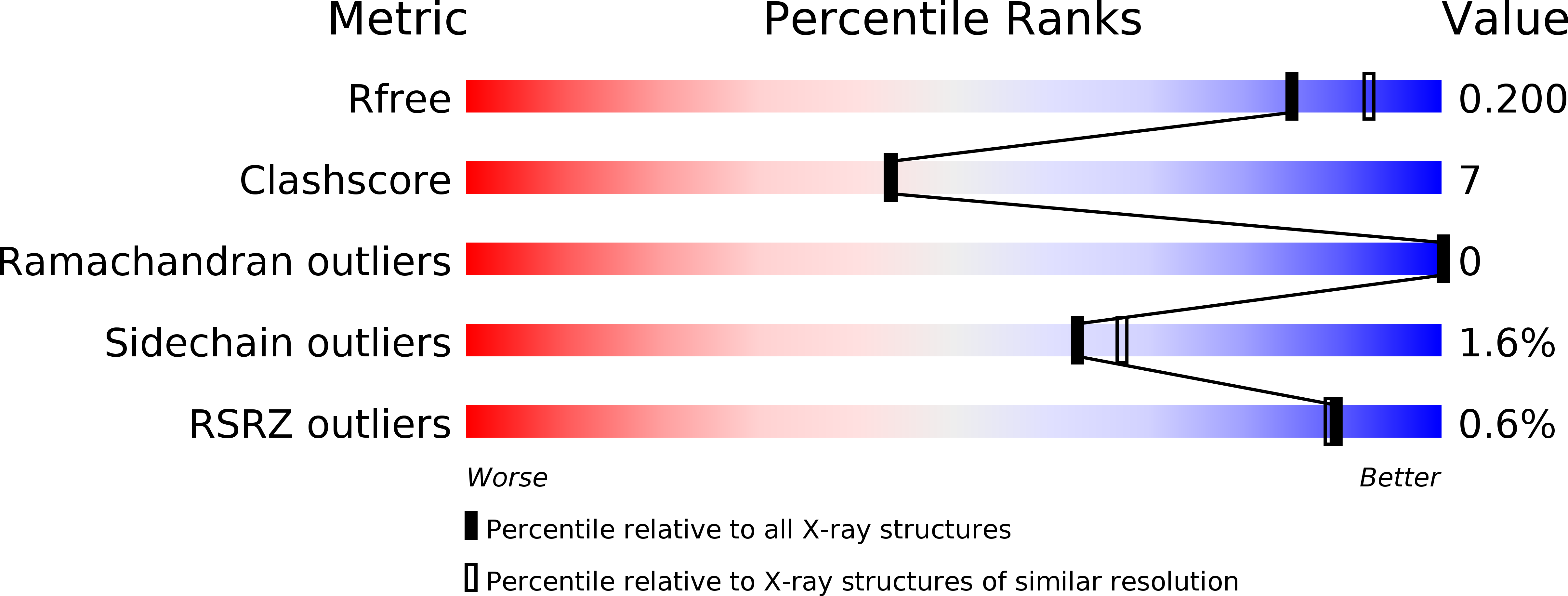
1WP4 image
Deposition Date 2004-08-30
Release Date 2005-08-30
Last Version Date 2023-10-25
Entry Detail
PDB ID:
1WP4
Keywords:
Title:
Structure of TT368 protein from Thermus Thermophilus HB8
Biological Source:
Source Organism:
Method Details:
Experimental Method:
Resolution:
2.00 Å
R-Value Free:
0.20
R-Value Work:
0.18
R-Value Observed:
0.18
Space Group:
P 21 21 21
Macromolecular Entities
Polymer Type:polypeptide(L)
Molecule:3-hydroxyisobutyrate dehydrogenase
Gene (Uniprot):TTHA0237
Chain IDs:A, B, C, D
Chain Length:289
Number of Molecules:4
Biological Source:Thermus thermophilus
Primary Citation
Crystal Structure of Novel NADP-dependent 3-Hydroxyisobutyrate Dehydrogenase from Thermus thermophilus HB8
J.Mol.Biol. 352 905 917 (2005)
PMID: 16126223 DOI: 10.1016/j.jmb.2005.07.068

Abstact

3-Hydroxyisobutyrate, a central metabolite in the valine catabolic pathway, is reversibly oxidized to methylmalonate semialdehyde by a specific dehydrogenase belonging to the 3-hydroxyacid dehydrogenase family. To gain insight into the function of this enzyme at the atomic level, we have determined the first crystal structures of the 3-hydroxyisobutyrate dehydrogenase from Thermus thermophilus HB8: holo enzyme and sulfate ion complex. The crystal structures reveal a unique tetrameric oligomerization and a bound cofactor NADP+. This bacterial enzyme may adopt a novel cofactor-dependence on NADP, whereas NAD is preferred in eukaryotic enzymes. The protomer folds into two distinct domains with open/closed interdomain conformations. The cofactor NADP+ with syn nicotinamide and the sulfate ion are bound to distinct sites located at the interdomain cleft of the protomer through an induced-fit domain closure upon cofactor binding. From the structural comparison with the crystal structure of 6-phosphogluconate dehydrogenase, another member of the 3-hydroxyacid dehydrogenase family, it is suggested that the observed sulfate ion and the substrate 3-hydroxyisobutyrate share the same binding pocket. The observed oligomeric state might be important for the catalytic function through forming the active site involving two adjacent subunits, which seems to be conserved in the 3-hydroxyacid dehydrogenases. A kinetic study confirms that this enzyme has strict substrate specificity for 3-hydroxyisobutyrate and serine, but it cannot distinguish the chirality of the substrates. Lys165 is likely the catalytic residue of the enzyme.

Legend

Protein

Chemical

Disease

Primary Citation of related structures