1WNH image
Deposition Date 2004-08-04
Release Date 2005-02-15
Last Version Date 2024-03-13
Entry Detail
PDB ID:
1WNH
Title:
Crystal structure of mouse Latexin (tissue carboxypeptidase inhibitor)
Biological Source:
Source Organism:
Mus musculus (Taxon ID: 10090)
Host Organism:
Method Details:
Experimental Method:
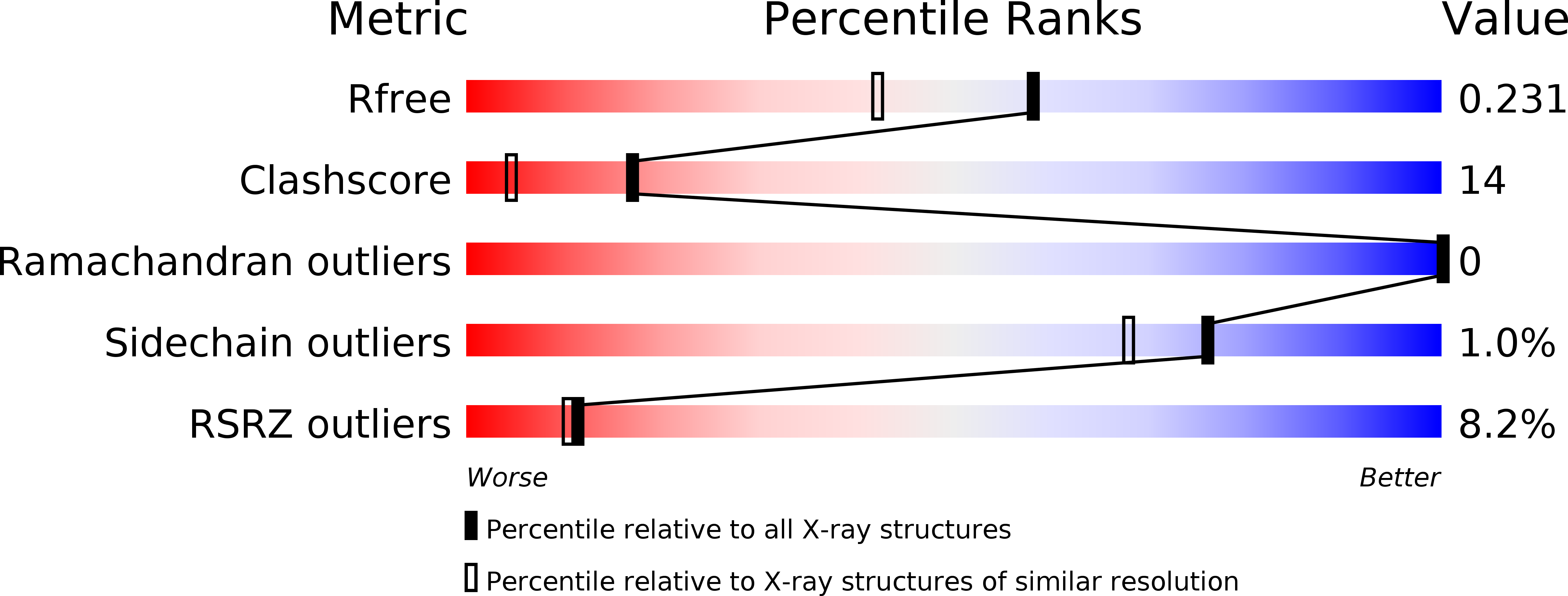
Resolution:
1.83 Å
R-Value Free:
0.23
R-Value Work:
0.21
R-Value Observed:
0.21
Space Group:
P 43 21 2
Macromolecular Entities
Polymer Type:polypeptide(L)
Molecule:Latexin
Gene (Uniprot):Lxn
Chain IDs:A
Chain Length:225
Number of Molecules:1
Biological Source:Mus musculus
Primary Citation
An Inflammatory Role for the Mammalian Carboxypeptidase Inhibitor Latexin: Relationship to Cystatins and the Tumor Suppressor TIG1
Structure 13 309 317 (2005)
PMID: 15698574 DOI: 10.1016/j.str.2004.12.013

Abstact

Latexin, the only known mammalian carboxypeptidase inhibitor, has no detectable sequence similarity with plant and parasite inhibitors, but it is related to a human putative tumor suppressor protein, TIG1. Latexin is expressed in the developing brain, and we find that it plays a role in inflammation, as it is expressed at high levels and is inducible in macrophages in concert with other protease inhibitors and potential protease targets. The crystal structure of mouse latexin, solved at 1.83 A resolution, shows no structural relationship with other carboxypeptidase inhibitors. Furthermore, despite a lack of detectable sequence duplication, the structure incorporates two topologically analogous domains related by pseudo two-fold symmetry. Surprisingly, these domains share a cystatin fold architecture found in proteins that inhibit cysteine proteases, suggesting an evolutionary and possibly functional relationship. The structure of the tumor suppressor protein TIG1 was modeled, revealing its putative membrane binding surface.

Legend

Protein

Chemical

Disease

Primary Citation of related structures