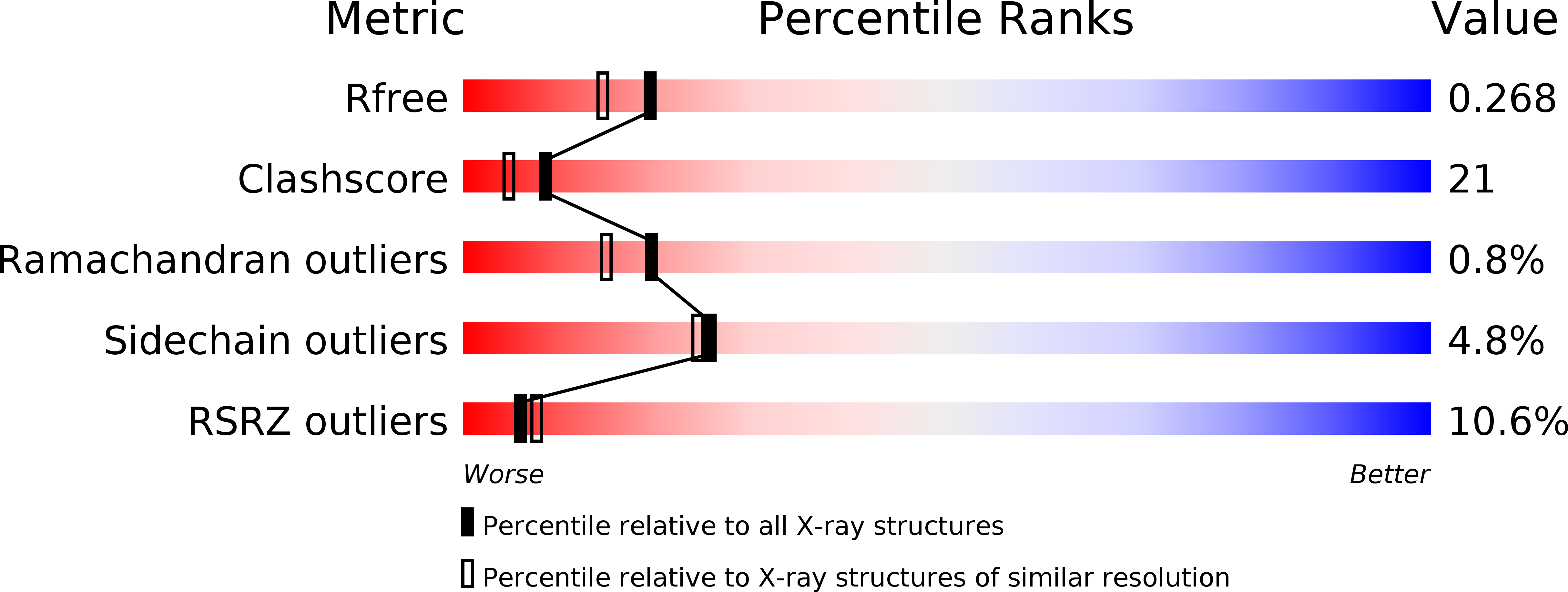
1WMG image
Deposition Date 2004-07-09
Release Date 2005-01-09
Last Version Date 2024-11-20
Entry Detail
PDB ID:
1WMG
Keywords:
Title:
Crystal structure of the UNC5H2 death domain
Biological Source:
Source Organism:
Mus musculus (Taxon ID: 10090)
Method Details:
Experimental Method:
Resolution:
2.10 Å
R-Value Free:
0.26
R-Value Work:
0.22
R-Value Observed:
0.22
Space Group:
C 1 2 1
Macromolecular Entities
Structures with similar UniProt ID
Protein Blast
Polymer Type:polypeptide(L)
Molecule:netrin receptor Unc5h2
Gene (Uniprot):Unc5b
Chain IDs:A, B, C, D, E, F
Chain Length:103
Number of Molecules:6
Biological Source:Mus musculus
Modified Residue
Compound ID Chain ID Parent Comp ID Details 2D Image
MSE A MET SELENOMETHIONINE
Primary Citation
Structure of the UNC5H2 death domain
ACTA CRYSTALLOGR.,SECT.D 62 1502 1509 (2006)
PMID: 17139086 DOI: 10.1107/S0907444906039369

Abstact

UNC5Hs (UNC5H1-4) are netrin 1 receptors that are involved in axonal guidance and neuronal migration. They are dependence receptors that mediate apoptosis in the absence of netrin 1. UNC5H2-induced apoptosis depends on the interaction of the death domain at the C-terminus with the DAP-kinase death domain and caspase cleavage near the transmembrane region. Here, the crystal structure of the mouse UNC5H2 death domain has been determined at 2.1 A resolution. The domain adopts a six-helix bundle fold, which is similar to those of the other members of the death-domain superfamily. The UNC5H2 death domain is a dimer in the crystal and in solution. This homodimerized structure may represent the structure of the death domain when netrin 1 binds to the UNC5H2 receptor. Homodimerization of UNC5H2 may block the access of caspase to the cleavage site. In the death-domain dimer, residues in alpha3 and the 3(10)-helix preceding alpha3 and the residues in alpha4 make significant contacts, mainly by hydrophobic and van der Waals interactions.

Legend

Protein

Chemical

Disease

Primary Citation of related structures