Deposition Date
2004-07-03
Release Date
2004-11-02
Last Version Date
2024-05-29
Entry Detail
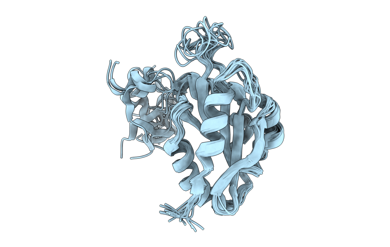
PDB ID:
1WM4
Keywords:
Title:
Solution structure of mouse coactosin, an actin filament binding protein
Biological Source:
Source Organism(s):
Mus musculus (Taxon ID: 10090)
Expression System(s):
Method Details:
Experimental Method:
Conformers Calculated:
100
Conformers Submitted:
15
Selection Criteria:
structures with the lowest energy