1WKY image
Deposition Date 2004-06-15
Release Date 2005-06-15
Last Version Date 2023-10-25
Entry Detail
PDB ID:
1WKY
Keywords:
Title:
Crystal structure of alkaline mannanase from Bacillus sp. strain JAMB-602: catalytic domain and its Carbohydrate Binding Module
Biological Source:
Source Organism:
Bacillus sp. (Taxon ID: 244966)
Host Organism:
Method Details:
Experimental Method:
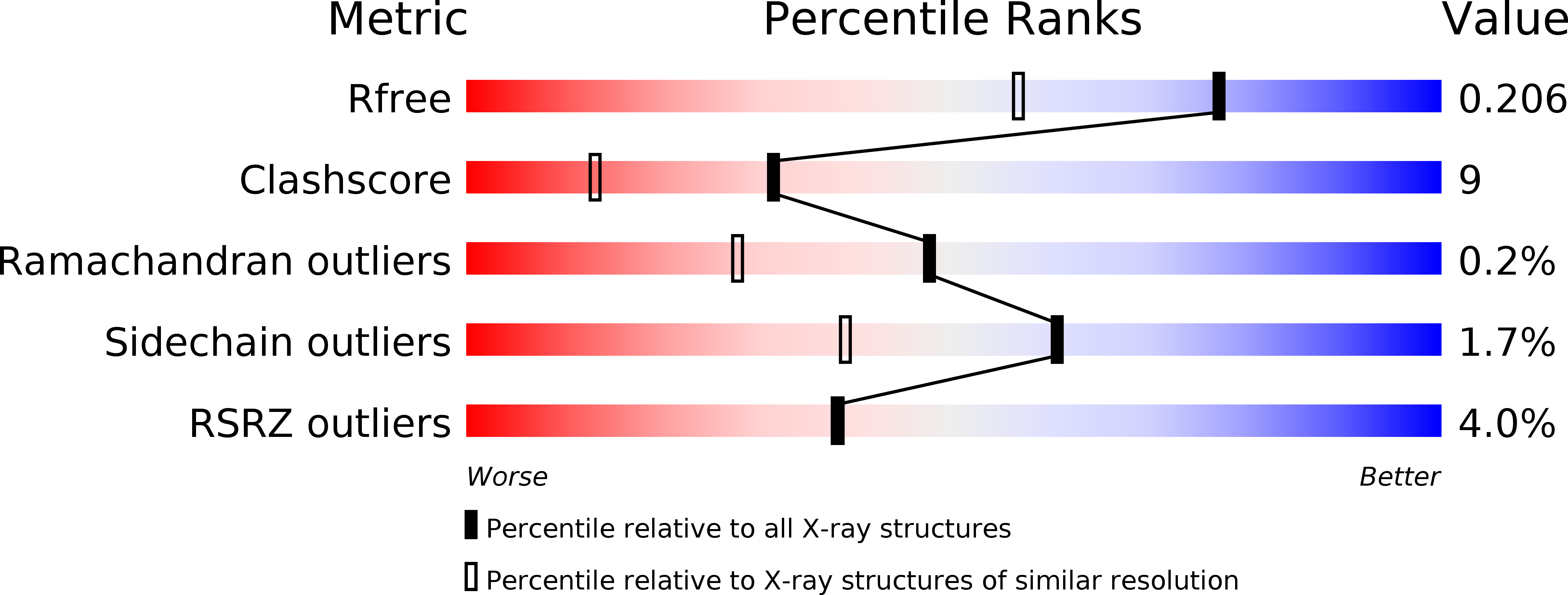
Resolution:
1.65 Å
R-Value Free:
0.2
R-Value Work:
0.17
R-Value Observed:
0.17
Space Group:
P 21 21 21
Macromolecular Entities
Structures with similar UniProt ID
Protein Blast
Polymer Type:polypeptide(L)
Molecule:endo-beta-1,4-Mannanase
Gene (Uniprot):amn5A
Chain IDs:A
Chain Length:464
Number of Molecules:1
Biological Source:Bacillus sp.
Primary Citation
Crystallization and preliminary X-ray study of alkaline mannanase from an alkaliphilic Bacillus isolate.
Acta Crystallogr.,Sect.D 60 1490 1492 (2004)
PMID: 15272186 DOI: 10.1107/S0907444904014313

Abstact

An alkaline mannanase (EC 3.2.1.78) from the alkaliphilic Bacillus sp. strain JAMB-602 was cloned and sequenced. The deduced amino-acid sequence of the enzyme suggested that the enzyme consists of a catalytic and unknown additional domains. The recombinant enzyme expressed by B. subtilis was crystallized using the hanging-drop vapour-diffusion method at 277 K. X-ray diffraction data were collected to 1.65 A. The crystal belongs to space group P2(1)2(1)2(1), with unit-cell parameters a = 70.7, b = 79.5, c = 80.4 A. The asymmetric unit contains one protein molecule, with a corresponding VM of 2.26 A3 Da(-1) and a solvent content of 45.6%. Molecular replacement for initial phasing was carried out using the three-dimensional structure of a mannanase from Thermomonospora fusca as a search model, which corresponds to the catalytic domain of the alkaline mannanase. It gave sufficient phases to build the unknown domain.

Legend

Protein

Chemical

Disease

Primary Citation of related structures
Feedback Form
Name
Email
Institute
Feedback