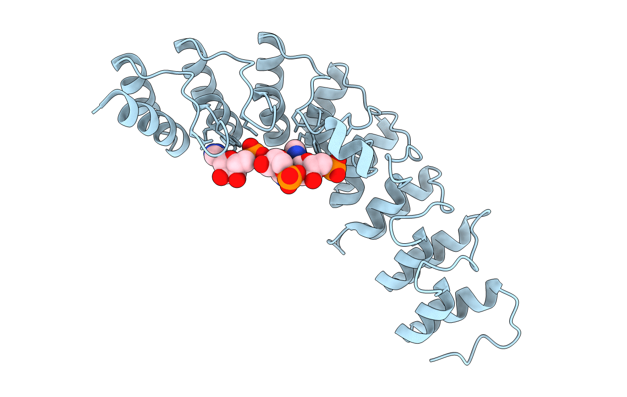
1WDY image
Entry Detail
PDB ID:
1WDY
Keywords:
Title:
Crystal structure of ribonuclease
Biological Source:
Source Organism:
Host Organism:
PDB Version:
Deposition Date:
2004-05-19
Release Date:
2004-10-05
Method Details:
Experimental Method:
Resolution:
1.80 Å
R-Value Free:
0.22
R-Value Work:
0.20
R-Value Observed:
0.20
Space Group:
P 21 21 21
Macromolecular Entities
Polymer Type:polypeptide(L)
Description:2-5A-dependent ribonuclease
Mutations:I97K
Chain IDs:A
Chain Length:285
Number of Molecules:1
Biological Source:Homo sapiens
Ligand Molecules
Primary Citation
Structural basis for recognition of 2',5'-linked oligoadenylates by human ribonuclease L
Embo J. 23 3929 3938 (2004)
PMID: 15385955 DOI: 10.1038/sj.emboj.7600420

Abstact

An interferon-induced endoribonuclease, ribonuclease L (RNase L), is implicated in both the molecular mechanism of action of interferon and the fundamental control of RNA stability in mammalian cells. RNase L is catalytically active only after binding to an unusual activator molecule containing a 5'-phosphorylated 2',5'-linked oligoadenylate (2-5A), in the N-terminal half. Here, we report the crystal structure of the N-terminal ankyrin repeat domain (ANK) of human RNase L complexed with the activator 2-5A. This is the first structural view of an ankyrin repeat structure directly interacting with a nucleic acid, rather than with a protein. The ANK domain folds into eight ankyrin repeat elements and forms an extended curved structure with a concave surface. The 2-5A molecule is accommodated at a concave site and directly interacts with ankyrin repeats 2-4. Interestingly, two structurally equivalent 2-5A binding motifs are found at repeats 2 and 4. The structural basis for 2-5A recognition by ANK is essential for designing stable 2-5As with a high likelihood of activating RNase L.

Legend

Protein

Chemical

Disease

Primary Citation of related structures