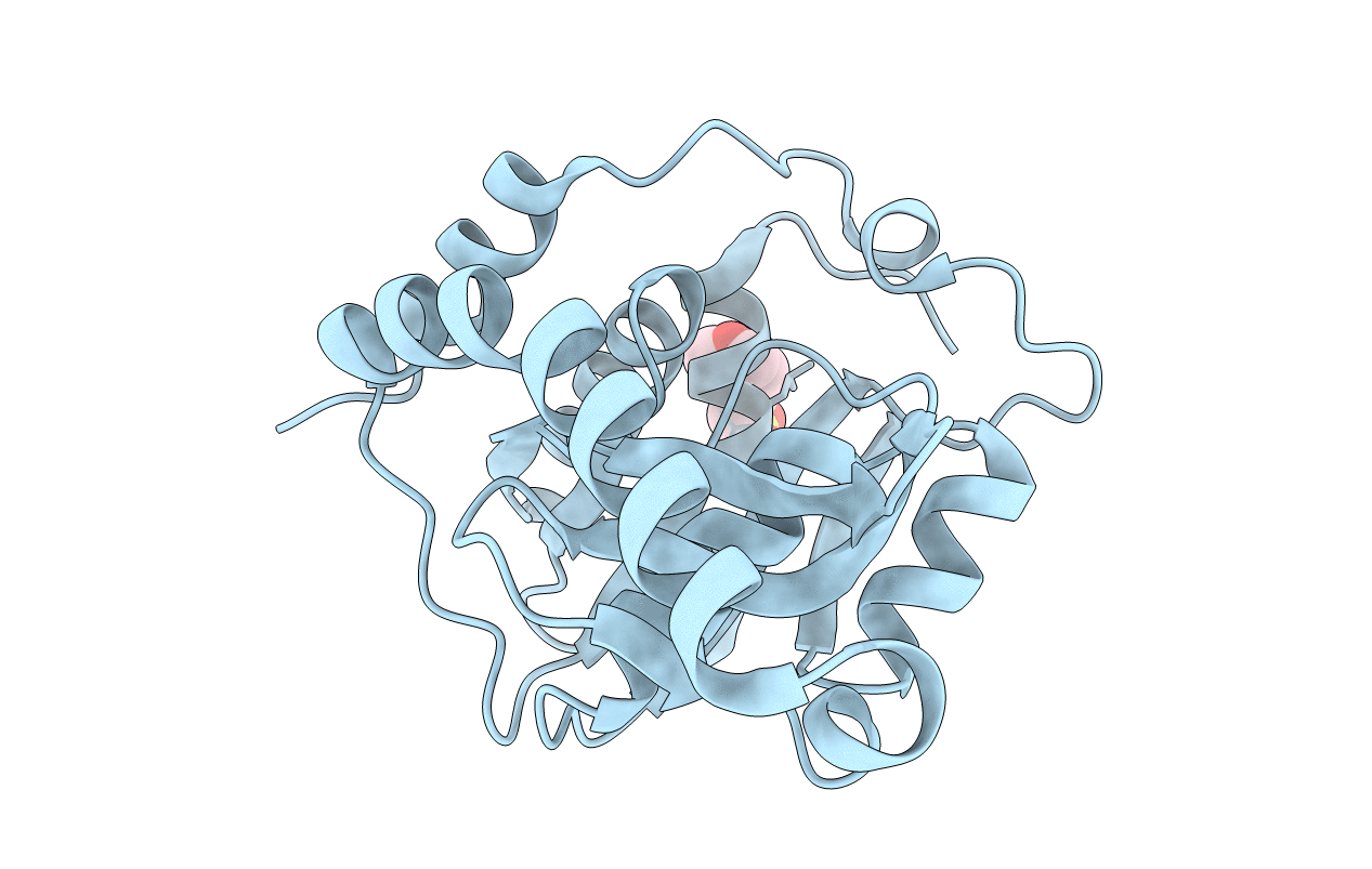
1WD5 image
Deposition Date 2004-05-11
Release Date 2004-11-11
Last Version Date 2024-11-06
Entry Detail
PDB ID:
1WD5
Title:
Crystal structure of TT1426 from Thermus thermophilus HB8
Biological Source:
Source Organism:
Host Organism:
Method Details:
Experimental Method:
Resolution:
2.00 Å
R-Value Free:
0.22
R-Value Work:
0.19
R-Value Observed:
0.19
Space Group:
P 41 21 2
Macromolecular Entities
Structures with similar UniProt ID
Protein Blast
Polymer Type:polypeptide(L)
Molecule:hypothetical protein TT1426
Gene (Uniprot):TTHA1462
Chain IDs:A
Chain Length:208
Number of Molecules:1
Biological Source:Thermus thermophilus
Modified Residue
Compound ID Chain ID Parent Comp ID Details 2D Image
MSE A MET SELENOMETHIONINE
Ligand Molecules
Primary Citation
Crystal structure of a predicted phosphoribosyltransferase (TT1426) from Thermus thermophilus HB8 at 2.01 A resolution
Protein Sci. 14 823 827 (2005)
PMID: 15689504 DOI: 10.1110/ps.041229405

Abstact

TT1426, from Thermus thermophilus HB8, is a conserved hypothetical protein with a predicted phosphoribosyltransferase (PRTase) domain, as revealed by a Pfam database search. The 2.01 A crystal structure of TT1426 has been determined by the multiwavelength anomalous dispersion (MAD) method. TT1426 comprises a core domain consisting of a central five-stranded beta sheet surrounded by four alpha-helices, and a subdomain in the C terminus. The core domain structure resembles those of the type I PRTase family proteins, although a significant structural difference exists in an inserted 43-residue region. The C-terminal subdomain corresponds to the "hood," which contains a substrate-binding site in the type I PRTases. The hood structure of TT1426 differs from those of the other type I PRTases, suggesting the possibility that TT1426 binds an unknown substrate. The structure-based sequence alignment provides clues about the amino acid residues involved in catalysis and substrate binding.

Legend

Protein

Chemical

Disease

Primary Citation of related structures