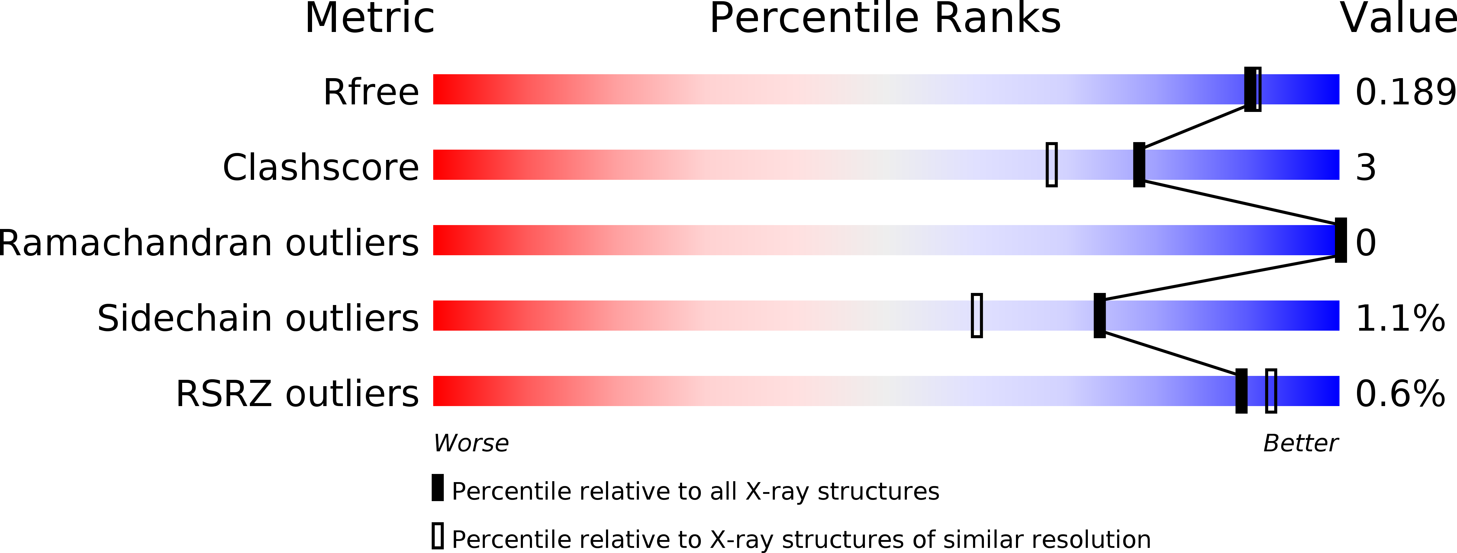
1WD3 image
Deposition Date 2004-05-11
Release Date 2004-09-14
Last Version Date 2024-10-16
Entry Detail
PDB ID:
1WD3
Keywords:
Title:
Crystal structure of arabinofuranosidase
Biological Source:
Source Organism:
Host Organism:
Method Details:
Experimental Method:
Resolution:
1.75 Å
R-Value Free:
0.20
R-Value Work:
0.19
R-Value Observed:
0.19
Space Group:
P 21 21 21
Macromolecular Entities
Polymer Type:polypeptide(L)
Molecule:alpha-L-arabinofuranosidase B
Chain IDs:A
Chain Length:482
Number of Molecules:1
Biological Source:Aspergillus kawachii
Modified Residue
Compound ID Chain ID Parent Comp ID Details 2D Image
ASN A ASN GLYCOSYLATION SITE
Primary Citation
Crystal structure of a family 54 alpha-L-arabinofuranosidase reveals a novel carbohydrate-binding module that can bind arabinose
J.Biol.Chem. 279 44907 44914 (2004)
PMID: 15292273 DOI: 10.1074/jbc.M405390200

Abstact

As the first known structures of a glycoside hydrolase family 54 (GH54) enzyme, we determined the crystal structures of free and arabinose-complex forms of Aspergillus kawachii IFO4308 alpha-l-arabinofuranosidase (AkAbfB). AkAbfB comprises two domains: a catalytic domain and an arabinose-binding domain (ABD). The catalytic domain has a beta-sandwich fold similar to those of clan-B glycoside hydrolases. ABD has a beta-trefoil fold similar to that of carbohydrate-binding module (CBM) family 13. However, ABD shows a number of characteristics distinctive from those of CBM family 13, suggesting that it could be classified into a new CBM family. In the arabinose-complex structure, one of three arabinofuranose molecules is bound to the catalytic domain through many interactions. Interestingly, a disulfide bond formed between two adjacent cysteine residues recognized the arabinofuranose molecule in the active site. From the location of this arabinofuranose and the results of a mutational study, the nucleophile and acid/base residues were determined to be Glu(221) and Asp(297), respectively. The other two arabinofuranose molecules are bound to ABD. The O-1 atoms of the two arabinofuranose molecules bound at ABD are both pointed toward the solvent, indicating that these sites can both accommodate an arabinofuranose side-chain moiety linked to decorated arabinoxylans.

Legend

Protein

Chemical

Disease

Primary Citation of related structures