1WCF image
Deposition Date 2004-11-13
Release Date 2006-07-12
Last Version Date 2023-12-13
Entry Detail
PDB ID:
1WCF
Keywords:
Title:
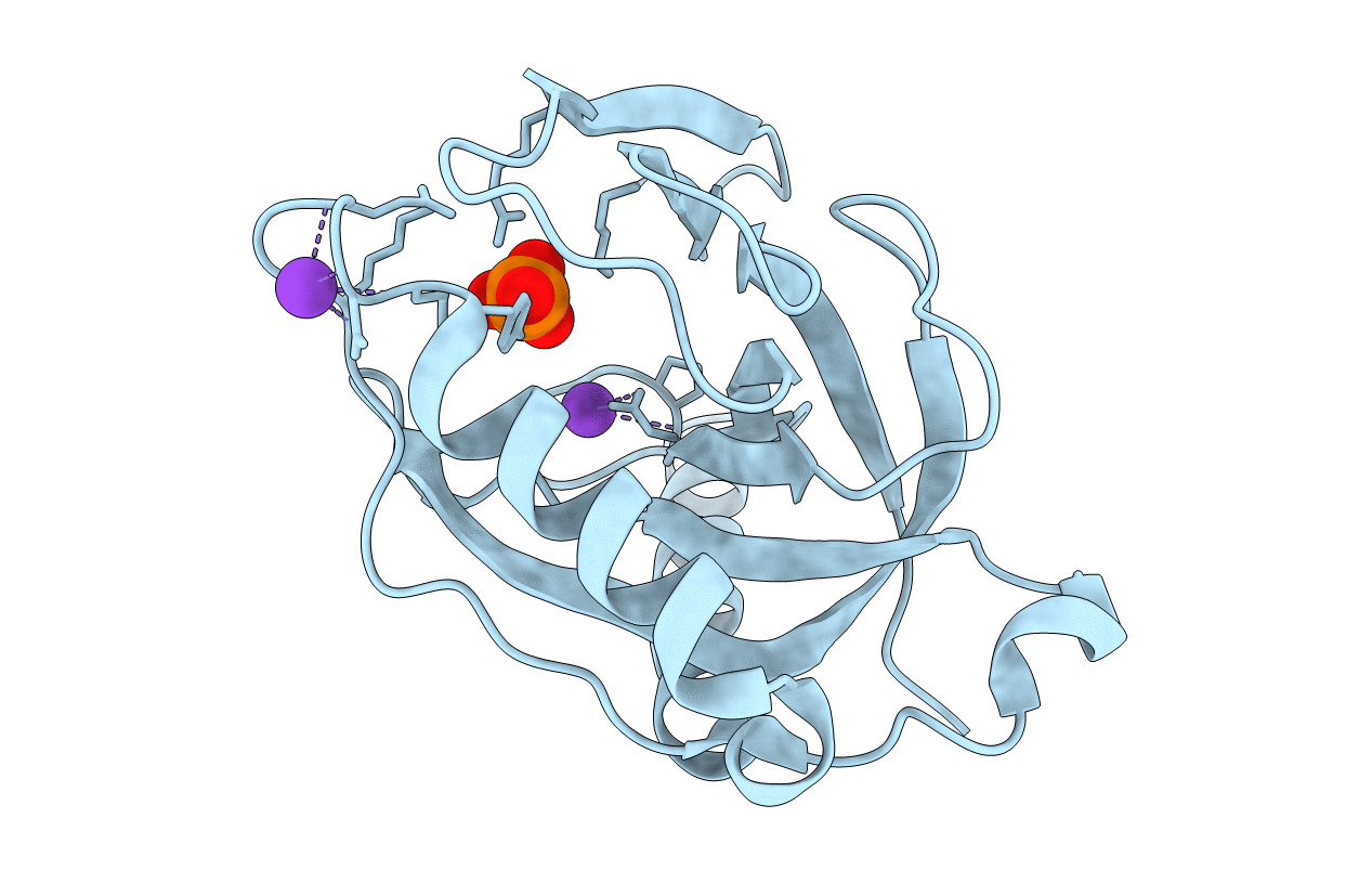
1.54 A CRYSTAL STRUCTURE OF RV3628, MYCOBACTERIUM TUBERCULOSIS INORGANIC PYROPHOSPHATASE (PPASE) AT PH7.0
Biological Source:
Source Organism:
Host Organism:
Method Details:
Experimental Method:
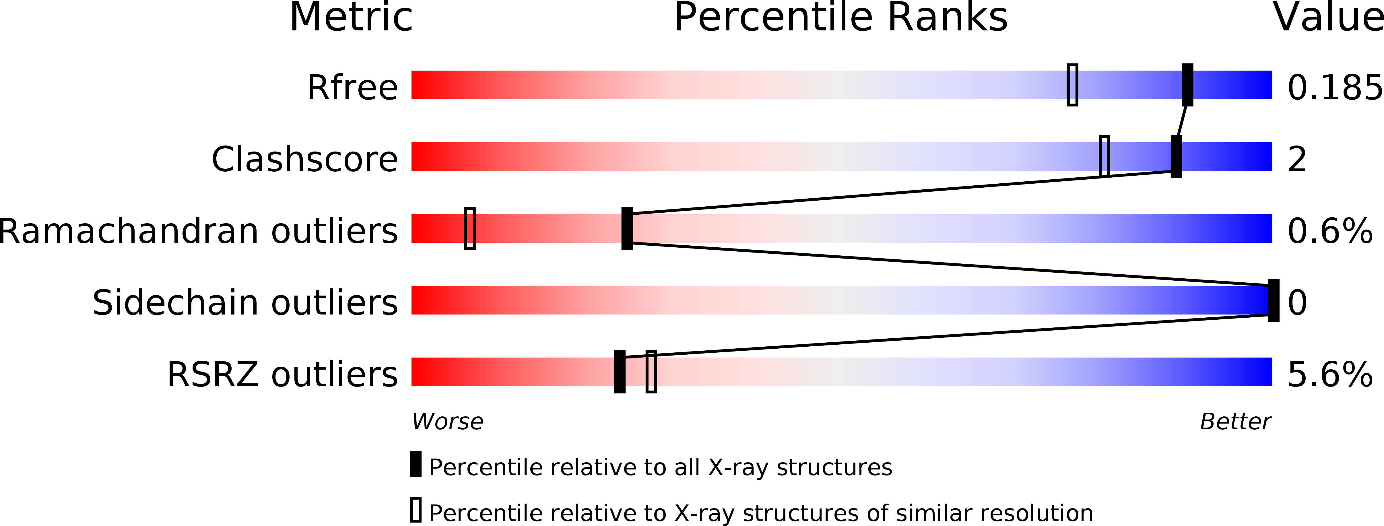
Resolution:
1.54 Å
R-Value Free:
0.17
R-Value Work:
0.15
R-Value Observed:
0.15
Space Group:
P 63 2 2
Macromolecular Entities
Structures with similar UniProt ID
Protein Blast
Polymer Type:polypeptide(L)
Molecule:INORGANIC PYROPHOSPHATASE
Chain IDs:A
Chain Length:171
Number of Molecules:1
Biological Source:MYCOBACTERIUM TUBERCULOSIS
Primary Citation
Structure of the Mycobacterium Tuberculosis Soluble Inorganic Pyrophosphatase Rv3628 at Ph 7.0.
Acta Crystallogr.,Sect.F 67 866 ? (2011)
PMID: 21821883 DOI: 10.1107/S1744309111023323

Abstact

The 1.5 Å resolution crystal structure of the Mycobacterium tuberculosis soluble inorganic pyrophosphatase Rv3628 at pH 7.0 is reported. The M. tuberculosis and M. leprae genomes include genes for the only two family I inorganic pyrophosphatases known to contain two histidines in the active site. The role of these two residues in catalysis is not fully understood. Mutational and functional studies of the M. tuberculosis enzyme showed that His21 and His86 are not essential for pyrophosphate hydrolysis, but are responsible for a shift in the optimal pH for the reaction compared with the Escherichia coli enzyme. Comparison with the structure previously reported at pH 5.0 provides further insight into the role of the two histidines. Two potassium-binding sites are found as a result of the high potassium concentration in the mother liquor.

Legend

Protein

Chemical

Disease

Primary Citation of related structures
Feedback Form
Name
Email
Institute
Feedback