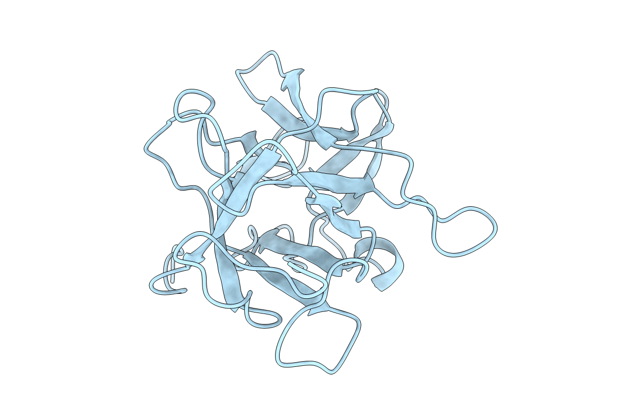
1WBC image
Deposition Date 1995-11-30
Release Date 1996-04-03
Last Version Date 2024-10-16
Entry Detail
PDB ID:
1WBC
Title:
CRYSTALLIZATION AND PRELIMINARY X-RAY STUDIES OF PSOPHOCARPIN B1, A CHYMOTRYPSIN INHIBITOR FROM WINGED BEAN SEEDS
Biological Source:
Source Organism:
Method Details:
Experimental Method:
Resolution:
2.95 Å
R-Value Work:
0.19
R-Value Observed:
0.19
Space Group:
P 61 2 2
Macromolecular Entities
Structures with similar UniProt ID
Protein Blast
Polymer Type:polypeptide(L)
Molecule:CHYMOTRYPSIN INHIBITOR (WCI)
Chain IDs:A
Chain Length:183
Number of Molecules:1
Biological Source:Psophocarpus tetragonolobus
Primary Citation
Structure of a Kunitz-type chymotrypsin from winged bean seeds at 2.95 A resolution.
Acta Crystallogr.,Sect.D 52 521 528 (1996)
PMID: 15299674 DOI: 10.1107/S0907444996000224

Abstact

Thc crystal structure of an alpha-chymotrypsin inhibitor (P6(1)22; a = 61.4, c = 210.9 A) isolated from winged bean (Psophocarpus. tetragonolobus) seeds has been determined at 2.95 A resolution by the molecular-replacement method using the 2.6 A coordinates of Erythrina trypsin inhibitor (ETI) as the starting model (57% sequence homology). This protease inhibitor, WCI, belongs to the Kunitz (STI) family and is a single polypeptide chain with 183 amino-acid residues having a molecular weight of 20 244 Da. Structure refinement with RESTRAIN and X-PLOR has led to a crystallographic R factor of 19.1% for 3469 observed reflections (I > 2sigma) in the resolution range 8-2.95 A. A total of 56 water molecules have been incorporated in the refined model containing 181 amino-acid residues. In the refined structure the deviations of bond lengths and bond angles from ideal values are 0.015 A and 2.2 degrees, respectively. The inhibitor molecule is spherical and consists of 12 antiparallel beta-strands with connecting loops arranged in a characteristic folding (a six-stranded beta-barrel and a six-stranded lid on one hollow end of the barrel) common to other homologous serine protease inhibitors in the Kunitz (STI) family as well as to some non-homologous proteins like interleukin-lalpha and interleukin-lbeta. In the structure the conformation of the protruding reactive-site loop is stabilized through hydrogen bonds mainly formed by the side chain of Asnl4, which intrudes inside the cavity of the reactive-site loop, with the side-chain and main-chain atoms of some residues in the loop region. A pseudo threefold axis exists parallel to the barrel axis of the structure. Each of the three subdomains comprises of four beta-strands with connecting loops.

Legend

Protein

Chemical

Disease

Primary Citation of related structures