1WAJ image
Deposition Date 1997-04-13
Release Date 1998-01-14
Last Version Date 2024-02-14
Entry Detail
PDB ID:
1WAJ
Title:
DNA POLYMERASE FROM BACTERIOPHAGE RB69
Biological Source:
Source Organism:
Host Organism:
Method Details:
Experimental Method:
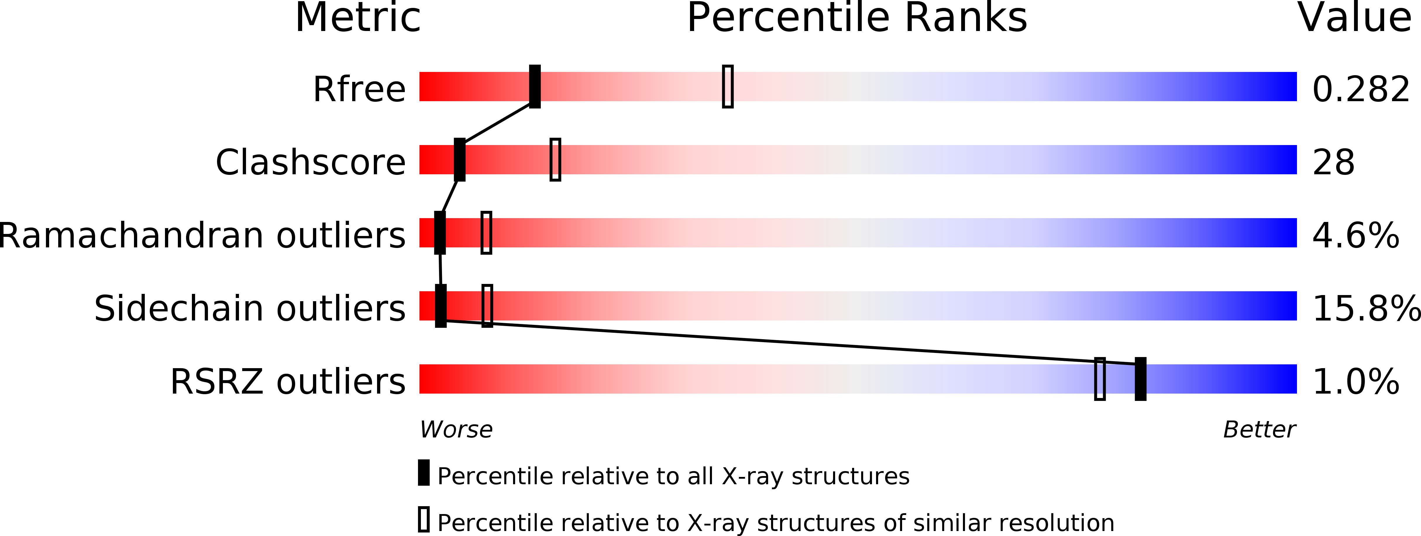
Resolution:
2.80 Å
R-Value Free:
0.28
R-Value Work:
0.21
R-Value Observed:
0.21
Space Group:
P 21 21 21
Macromolecular Entities
Structures with similar UniProt ID
Protein Blast
Polymer Type:polypeptide(L)
Molecule:DNA POLYMERASE
Gene (Uniprot):43
Chain IDs:A
Chain Length:903
Number of Molecules:1
Biological Source:Enterobacteria phage RB69
Ligand Molecules
Primary Citation
Crystal structure of a pol alpha family replication DNA polymerase from bacteriophage RB69.
Cell(Cambridge,Mass.) 89 1087 1099 (1997)
PMID: 9215631 DOI: 10.1016/S0092-8674(00)80296-2

Abstact

The 2.8 A resolution crystal structure of the bacteriophage RB69 gp43, a member of the eukaryotic pol alpha family of replicative DNA polymerases, shares some similarities with other polymerases but shows many differences. Although its palm domain has the same topology as other polymerases, except rat DNA polymerase beta, one of the three carboxylates required for nucleotidyl transfer is located on a different beta strand. The structures of the fingers and thumb domains are unrelated to all other known polymerase structures. The editing 3'-5' exonuclease domain of gp43 is homologous to that of E. coli DNA polymerase I but lies on the opposite side of the polymerase active site. An extended structure-based alignment of eukaryotic DNA polymerase sequences provides structural insights that should be applicable to most eukaryotic DNA polymerases.

Legend

Protein

Chemical

Disease

Primary Citation of related structures