1WA8 image
Deposition Date 2004-10-25
Release Date 2005-06-27
Last Version Date 2024-05-15
Entry Detail
PDB ID:
1WA8
Keywords:
Title:
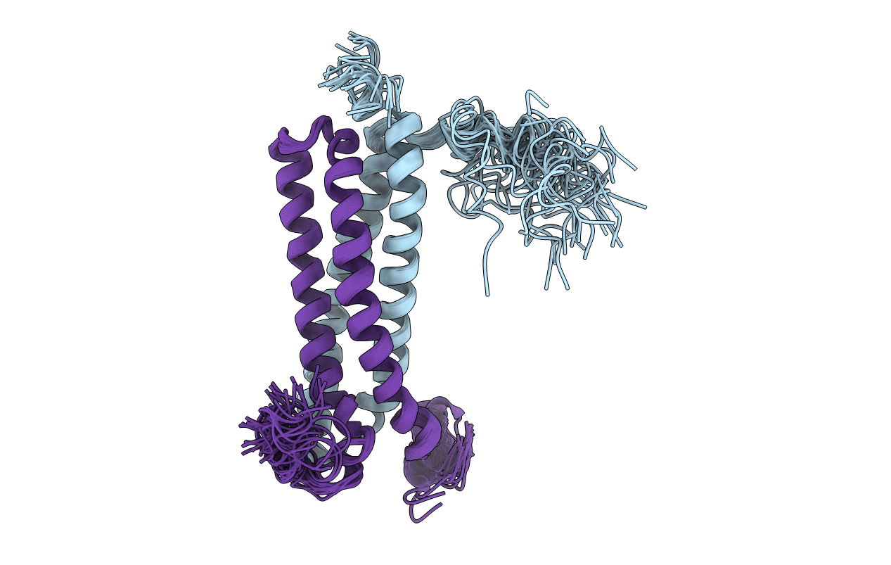
Solution Structure of the CFP-10.ESAT-6 Complex. Major Virulence Determinants of Pathogenic Mycobacteria
Biological Source:
Source Organism:
Host Organism:
Method Details:
Experimental Method:
Conformers Calculated:
100
Conformers Submitted:
28
Selection Criteria:
LEAST RESTRAINT VIOLATION
Macromolecular Entities
Structures with similar UniProt ID
Protein Blast
Polymer Type:polypeptide(L)
Molecule:ESAT-6 LIKE PROTEIN ESXB
Chain IDs:A
Chain Length:99
Number of Molecules:1
Biological Source:MYCOBACTERIUM BOVIS
Structures with similar UniProt ID
Protein Blast
Polymer Type:polypeptide(L)
Molecule:6 KDA EARLY SECRETORY ANTIGENIC TARGET (ESAT-6)
Chain IDs:B
Chain Length:95
Number of Molecules:1
Biological Source:MYCOBACTERIUM TUBERCULOSIS
Ligand Molecules
Primary Citation
Structure and Function of the Complex Formed by the Tuberculosis Virulence Factors Cfp-10 and Esat-6
Embo J. 24 2491 ? (2005)
PMID: 15973432 DOI: 10.1038/SJ.EMBOJ.7600732

Abstact

The secreted Mycobacterium tuberculosis complex proteins CFP-10 and ESAT-6 have recently been shown to play an essential role in tuberculosis pathogenesis. We have determined the solution structure of the tight, 1:1 complex formed by CFP-10 and ESAT-6, and employed fluorescence microscopy to demonstrate specific binding of the complex to the surface of macrophage and monocyte cells. A striking feature of the complex is the long flexible arm formed by the C-terminus of CFP-10, which was found to be essential for binding to the surface of cells. The surface features of the CFP-10.ESAT-6 complex, together with observed binding to specific host cells, strongly suggest a key signalling role for the complex, in which binding to cell surface receptors leads to modulation of host cell behaviour to the advantage of the pathogen.

Legend

Protein

Chemical

Disease

Primary Citation of related structures