1WA1 image
Deposition Date 2004-10-22
Release Date 2005-01-04
Last Version Date 2023-12-13
Entry Detail
PDB ID:
1WA1
Keywords:
Title:
Crystal Structure Of H313Q Mutant Of Alcaligenes Xylosoxidans Nitrite Reductase
Biological Source:
Source Organism:
Host Organism:
Method Details:
Experimental Method:
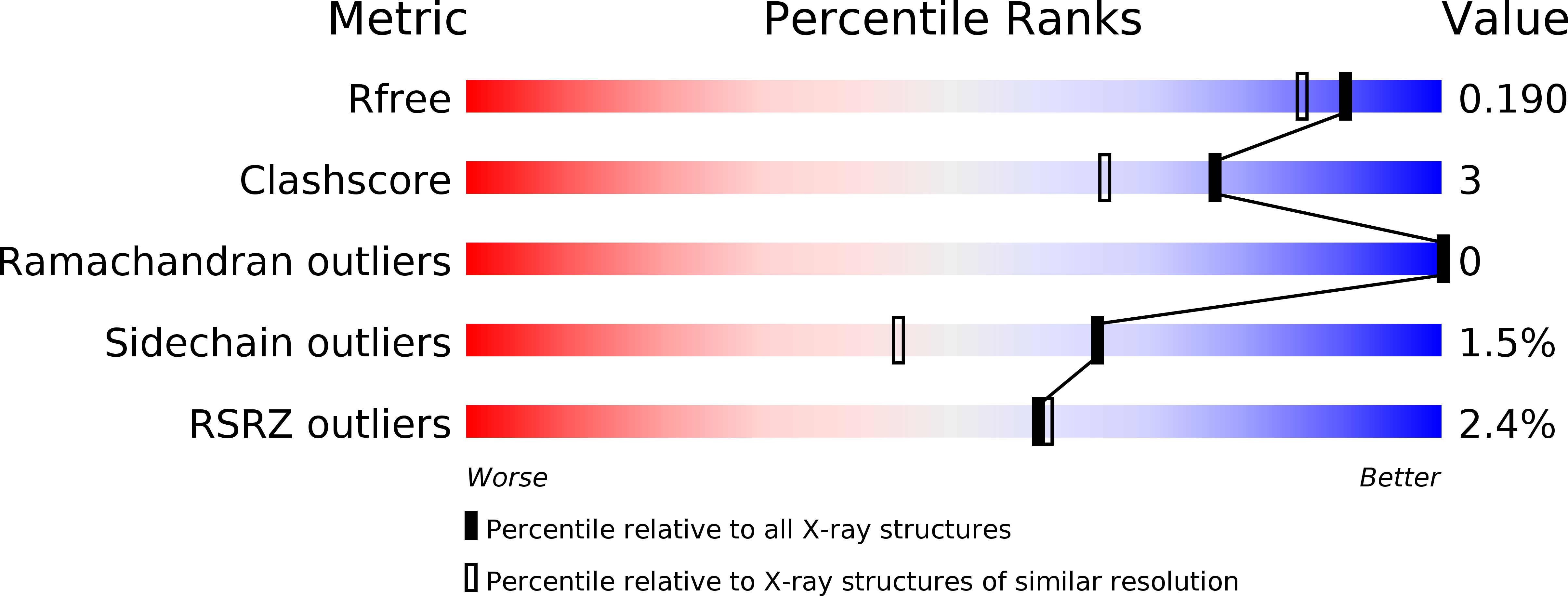
Resolution:
1.65 Å
R-Value Free:
0.19
R-Value Work:
0.16
R-Value Observed:
0.16
Space Group:
H 3
Macromolecular Entities
Polymer Type:polypeptide(L)
Molecule:DISSIMILATORY COPPER-CONTAINING NITRITE REDUCTASE
Gene (Uniprot):nir
Mutations:YES
Chain IDs:A (auth: X)
Chain Length:336
Number of Molecules:1
Biological Source:ALCALIGENES XYLOSOXIDANS
Primary Citation
Insights Into Redox Partner Interactions and Substrate Binding in Nitrite Reductase from Alcaligenes Xylosoxidans: Crystal Structures of the Trp138His and His313Gln Mutants
Biochemistry 43 16311 ? (2004)
PMID: 15610025 DOI: 10.1021/BI048682G

Abstact

Dissimilatory nitrite reductase catalyses the reduction of nitrite to nitric oxide within the key biological process of denitrification. We present biochemical and structural results on two key mutants, one postulated to be important for the interaction with the partner protein and the other for substrate entry. Trp138, adjacent to one of the type-1 Cu ligands, is one of the residues surrounding a small depression speculated to be important in complex formation with the physiological redox partners, azurin I and II. Our data reveal that the Trp138His mutant is fully active using methyl viologen as an artificial electron donor, but there is a large decrease in activity using azurin I. These observations together with its crystal structure at a high resolution of 1.6 A confirm the importance of Trp138 in electron transfer and thus in productive interaction with azurin. A "hydrophobic pocket" on the protein surface has been identified as the channel through which nitrite may be guided to the catalytic type-2 Cu site. Glu133 and His313 at the opening of the pocket are conserved among most blue and green copper nitrite reductases (CuNiRs). The failure to soak the substrate into our high-resolution crystal form of native and mutant CuNiRs has been linked to the observation of an extraneous poly(ethylene glycol) (PEG) molecule interacting with His313. We present the crystal structure of His313Gln and the substrate-bound mutant at high resolutions of 1.65 and 1.72 A, respectively. The observation of the substrate-bound structure for the His313Gln mutant and inhibitory studies with PEG establishes the role of the hydrophobic pocket as the port of substrate entry.

Legend

Protein

Chemical

Disease

Primary Citation of related structures