1W9Z image
Deposition Date 2004-10-21
Release Date 2005-04-01
Last Version Date 2024-05-08
Entry Detail
PDB ID:
1W9Z
Title:
Structure of Bannavirus VP9
Biological Source:
Source Organism:
BANNA VIRUS (Taxon ID: 77763)
Host Organism:
Method Details:
Experimental Method:
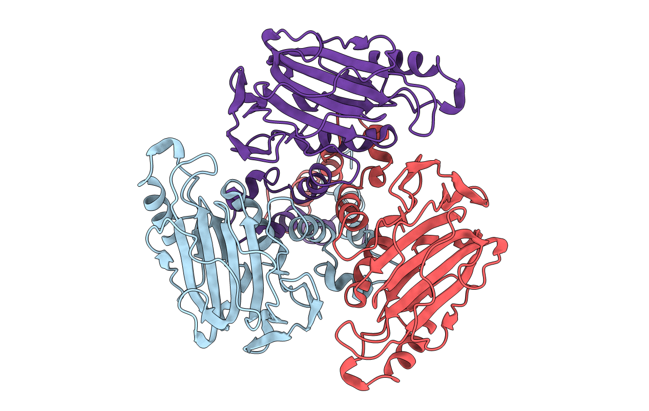
Resolution:
2.56 Å
R-Value Free:
0.24
R-Value Work:
0.18
R-Value Observed:
0.18
Space Group:
C 1 2 1
Macromolecular Entities
Polymer Type:polypeptide(L)
Molecule:VP9
Chain IDs:A, B, C
Chain Length:283
Number of Molecules:3
Biological Source:BANNA VIRUS
Primary Citation
The Structure and Function of the Outer Coat Protein Vp9 of Banna Virus
Structure 13 17 ? (2005)
PMID: 15642258 DOI: 10.1016/J.STR.2004.10.017

Abstact

Banna virus (BAV: genus Seadornavirus, family Reoviridae) has a double-shelled morphology similar to rotavirus and bluetongue virus. The structure of BAV outer-capsid protein VP9 was determined by X-ray crystallography at 2.6 A resolution, revealing a trimeric molecule, held together by an N-terminal helical bundle, reminiscent of coiled-coil structures found in fusion-active proteins such as HIV gp41. The major domain of VP9 contains stacked beta sheets with marked structural similarities to the receptor binding protein VP8 of rotavirus. Anti-VP9 antibodies neutralize viral infectivity, and, remarkably, pretreatment of cells with trimeric VP9 increased viral infectivity, indicating that VP9 is involved in virus attachment to cell surface and subsequent internalization. Sequence similarities were also detected between BAV VP10 and VP5 portion of rotavirus VP4, suggesting that the receptor binding and internalization apparatus, which is a single gene product activated by proteoloysis in rotavirus, is the product of two separate genome segments in BAV.

Legend

Protein

Chemical

Disease

Primary Citation of related structures