1W9S image
Deposition Date 2004-10-18
Release Date 2004-11-03
Last Version Date 2023-12-13
Entry Detail
PDB ID:
1W9S
Title:
Structure of a beta-1,3-glucan binding CBM6 from Bacillus halodurans
Biological Source:
Source Organism:
Host Organism:
Method Details:
Experimental Method:
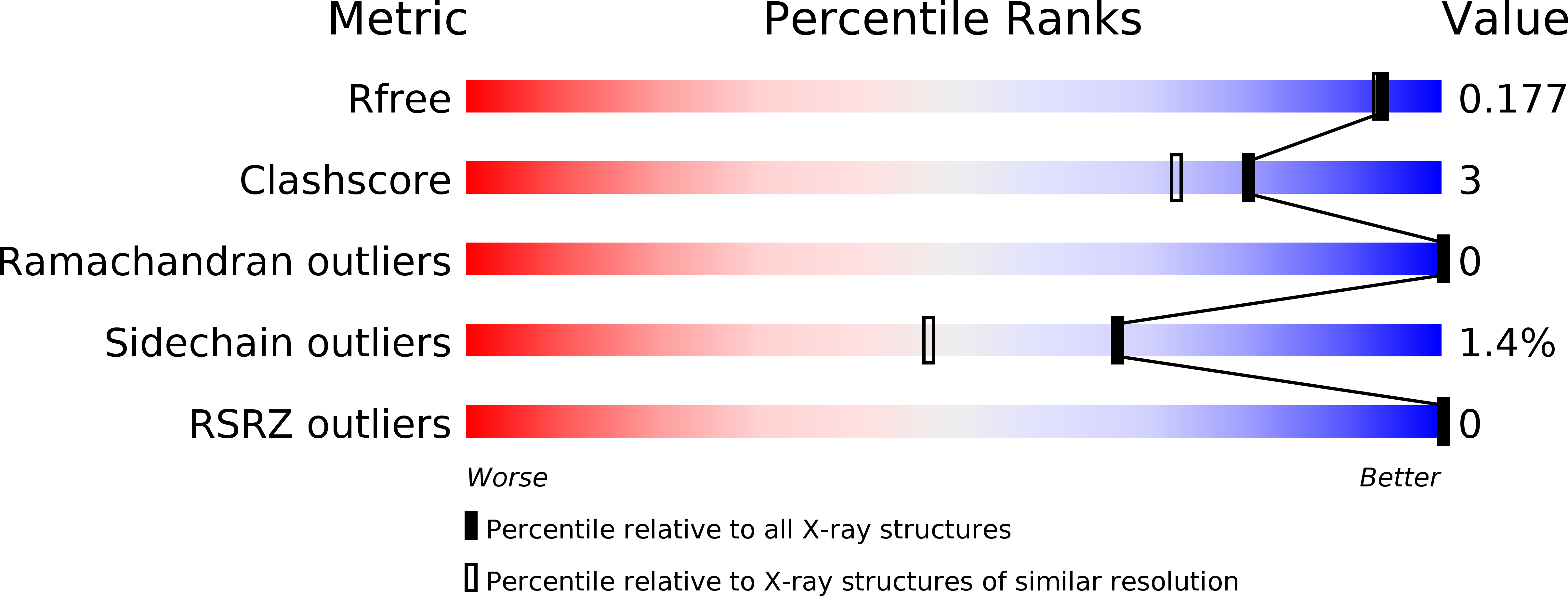
Resolution:
1.59 Å
R-Value Free:
0.16
R-Value Work:
0.11
R-Value Observed:
0.11
Space Group:
P 1
Macromolecular Entities
Polymer Type:polypeptide(L)
Molecule:BH0236 PROTEIN
Gene (Uniprot):BH0236
Chain IDs:A, B
Chain Length:142
Number of Molecules:2
Biological Source:BACILLUS HALODURANS
Primary Citation
Family 6 Carbohydrate Binding Modules Recognize the Non-Reducing End of Beta-1,3-Linked Glucans by Presenting a Unique Ligand Binding Surface
J.Biol.Chem. 280 530 ? (2005)
PMID: 15501830 DOI: 10.1074/JBC.M410113200

Abstact

Enzymes that hydrolyze insoluble complex polysaccharide structures contain non-catalytic carbohydrate binding modules (CBMS) that play a pivotal role in the action of these enzymes against recalcitrant substrates. Family 6 CBMs (CBM6s) are distinct from other CBM families in that these protein modules contain multiple distinct ligand binding sites, a feature that makes CBM6s particularly appropriate receptors for the beta-1,3-glucan laminarin, which displays an extended U-shaped conformation. To investigate the mechanism by which family 6 CBMs recognize laminarin, we report the biochemical and structural properties of a CBM6 (designated BhCBM6) that is located in an enzyme, which is shown, in this work, to display beta-1,3-glucanase activity. BhCBM6 binds beta-1,3-glucooligosaccharides with affinities of approximately 1 x 10(5) m(-1). The x-ray crystal structure of this CBM in complex with laminarihexaose reveals similarity with the structures of other CBM6s but a unique binding mode. The binding cleft in this protein is sealed at one end, which prevents binding of linear polysaccharides such as cellulose, and the orientation of the sugar at this site prevents glycone extension of the ligand and thus conferring specificity for the non-reducing ends of glycans. The high affinity for extended beta-1,3-glucooligosaccharides is conferred by interactions with the surface of the protein located between the two binding sites common to CBM6s and thus reveals a third ligand binding site in family 6 CBMs. This study therefore demonstrates how the multiple binding clefts and highly unusual protein surface of family 6 CBMs confers the extensive range of specificities displayed by this protein family. This is in sharp contrast to other families of CBMs where variation in specificity between different members reflects differences in the topology of a single binding site.

Legend

Protein

Chemical

Disease

Primary Citation of related structures