Deposition Date
2004-10-07
Release Date
2005-01-06
Last Version Date
2024-10-23
Entry Detail
PDB ID:
1W9A
Keywords:
Title:
Crystal structure of Rv1155 from Mycobacterium tuberculosis
Biological Source:
Source Organism:
MYCOBACTERIUM TUBERCULOSIS (Taxon ID: 83332)
Host Organism:
Method Details:
Experimental Method:
Resolution:
1.80 Å
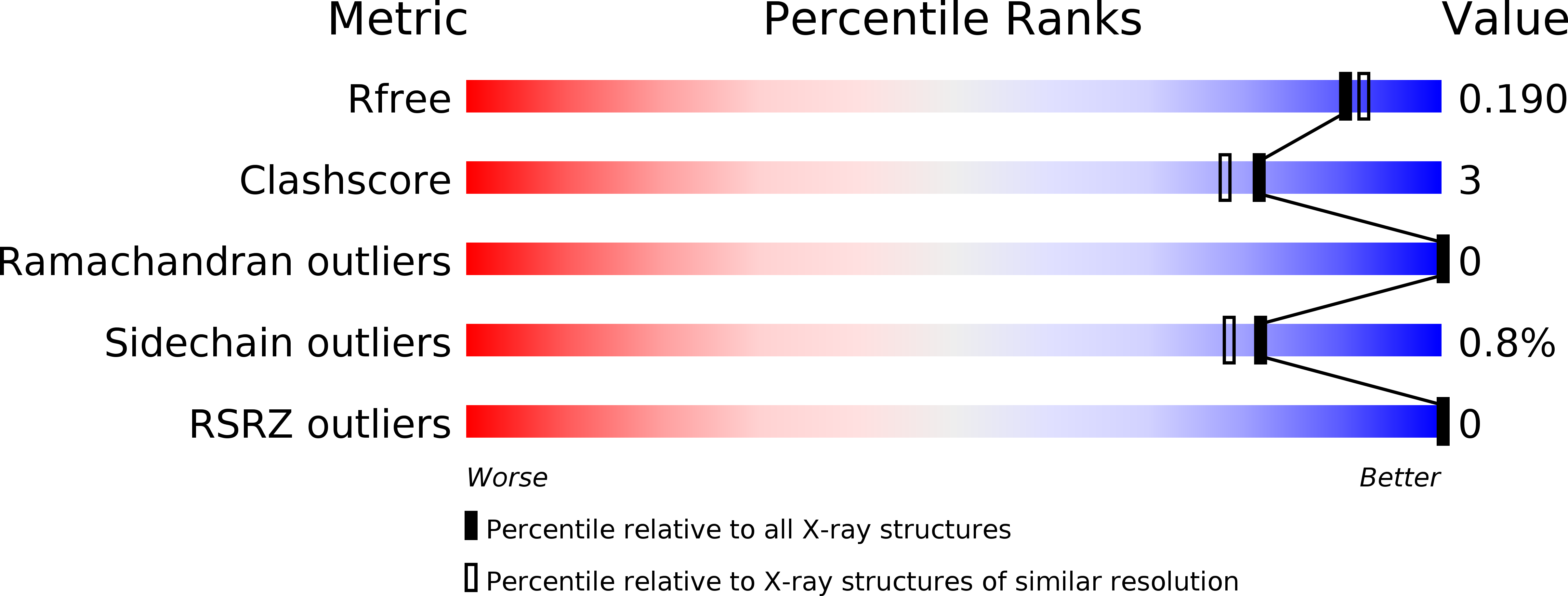
R-Value Free:
0.17
R-Value Work:
0.14
R-Value Observed:
0.14
Space Group:
P 1 21 1