Deposition Date
2004-09-20
Release Date
2006-08-30
Last Version Date
2024-10-16
Entry Detail
PDB ID:
1W8C
Keywords:
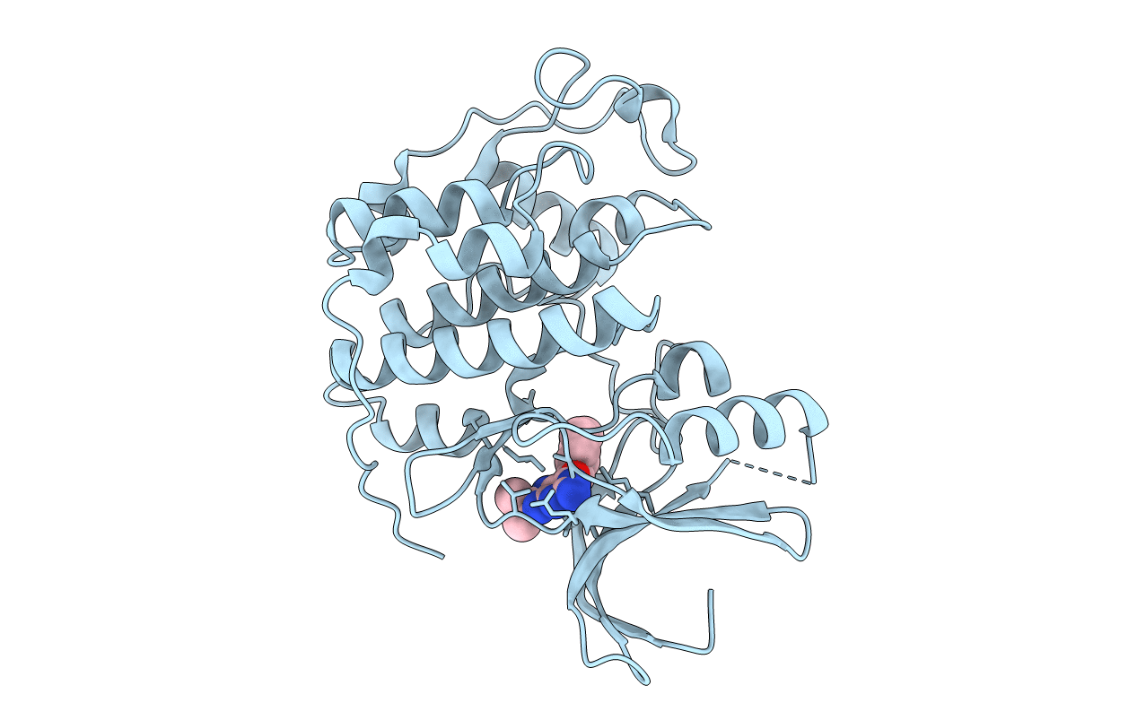
Title:
CO-CRYSTAL STRUCTURE OF 6-CYCLOHEXYLMETHOXY-8-ISOPROPYL-9H-PURIN-2- YLAMINE AND MONOMERIC CDK2
Biological Source:
Source Organism(s):
HOMO SAPIENS (Taxon ID: 9606)
Expression System(s):
Method Details:
Experimental Method:
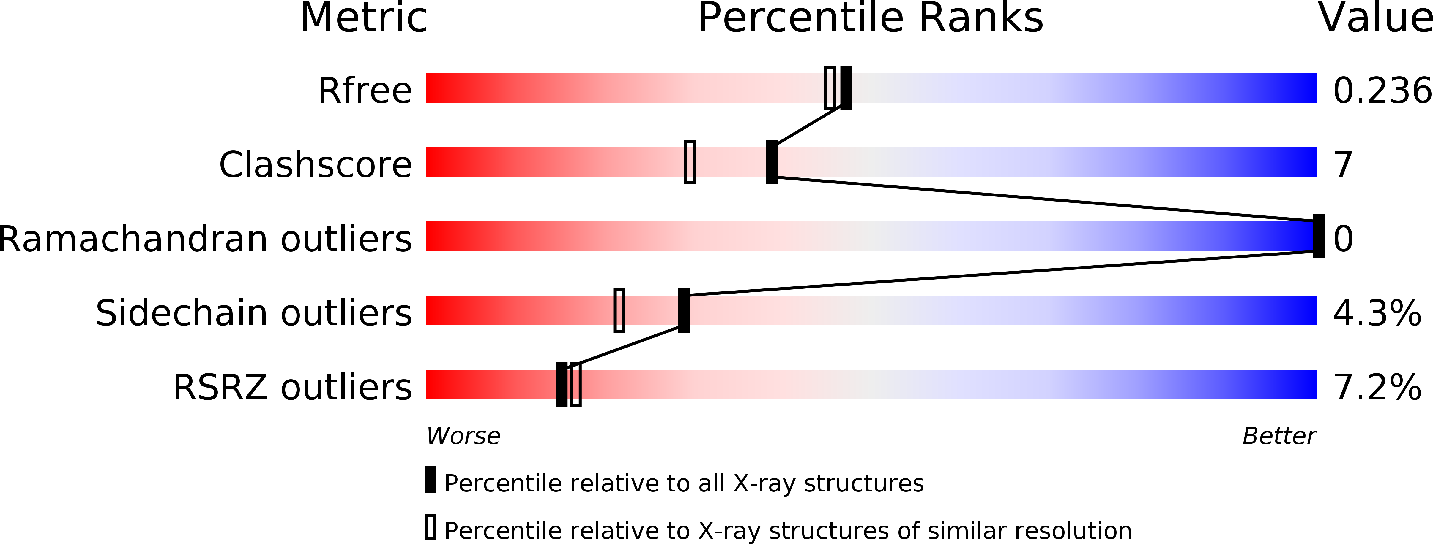
Resolution:
2.05 Å
R-Value Free:
0.25
R-Value Work:
0.19
R-Value Observed:
0.19
Space Group:
P 21 21 21