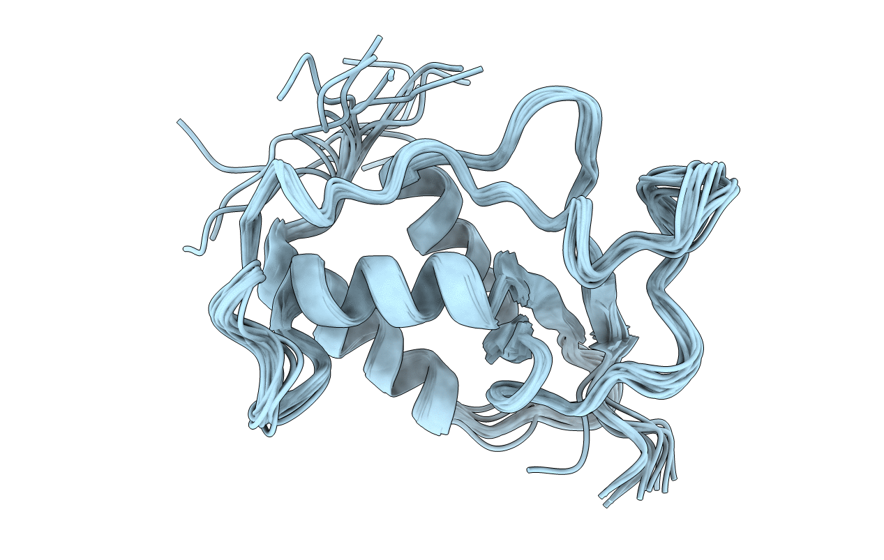
1W6V image
Deposition Date 2004-08-24
Release Date 2006-01-12
Last Version Date 2024-05-15
Entry Detail
PDB ID:
1W6V
Keywords:
Title:
Solution structure of the DUSP domain of hUSP15
Biological Source:
Source Organism:
HOMO SAPIENS (Taxon ID: 9606)
Host Organism:
Method Details:
Experimental Method:
Conformers Calculated:
20
Conformers Submitted:
13
Selection Criteria:
SMALLEST MAXIMUM RESTRAINT VIOLATION
Macromolecular Entities
Polymer Type:polypeptide(L)
Molecule:UBIQUITIN CARBOXYL-TERMINAL HYDROLASE 15
Gene (Uniprot):USP15
Chain IDs:A
Chain Length:141
Number of Molecules:1
Biological Source:HOMO SAPIENS
Ligand Molecules
Primary Citation
Solution Structure of the Human Ubiquitin-Specific Protease 15 Dusp Domain.
J.Biol.Chem. 281 5026 ? (2006)
PMID: 16298993 DOI: 10.1074/JBC.M510993200

Abstact

Ubiquitin-specific proteases (USPs) can remove covalently attached ubiquitin moieties from target proteins and regulate both the stability and ubiquitin-signaling state of their substrates. All USPs contain a conserved catalytic domain surrounded by one or more subdomains, some of which contribute to target recognition. One such specific subdomain, the DUSP domain (domain present in ubiquitin-specific proteases), is present in at least seven different human USPs that regulate the stability of or interact with the hypoxia-inducible transcription factor HIF1-alpha, the Von Hippel-Lindau protein (pVHL), cullin E3 ligases, and BRCA2. We describe the NMR solution structure of the DUSP domain of human USP15, recently implicated in COP9 (constitutive photomorphogenic gene 9)-signalosome regulation. Its tripod-like structure consists of a 3-fold alpha-helical bundle supporting a triple-stranded anti-parallel beta-sheet. The DUSP domain displays a novel fold, an alpha/beta tripod (AB3). DUSP domain surface properties and previously described work suggest a potential role in protein/protein interaction or substrate recognition.

Legend

Protein

Chemical

Disease

Primary Citation of related structures