1W6T image
Deposition Date 2004-08-24
Release Date 2005-08-22
Last Version Date 2023-12-13
Entry Detail
PDB ID:
1W6T
Keywords:
Title:
Crystal Structure Of Octameric Enolase From Streptococcus pneumoniae
Biological Source:
Source Organism:
Host Organism:
Method Details:
Experimental Method:
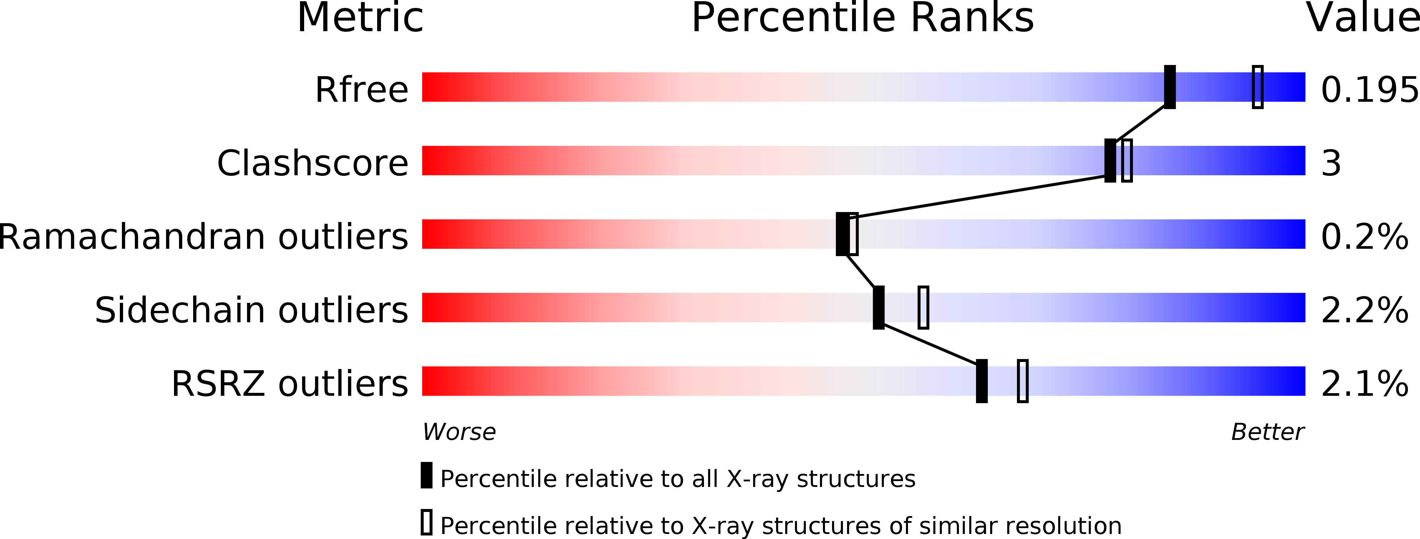
Resolution:
2.10 Å
R-Value Free:
0.18
R-Value Work:
0.14
R-Value Observed:
0.15
Space Group:
I 4
Macromolecular Entities
Polymer Type:polypeptide(L)
Molecule:ENOLASE
Chain IDs:A, B
Chain Length:444
Number of Molecules:2
Biological Source:STREPTOCOCCUS PNEUMONIAE
Primary Citation
Plasmin(Ogen)-Binding Alpha-Enolase from Streptococcus Pneumoniae: Crystal Structure and Evaluation of Plasmin(Ogen)-Binding Sites
J.Mol.Biol. 343 997 ? (2004)
PMID: 15476816 DOI: 10.1016/J.JMB.2004.08.088

Abstact

Alpha-enolases are ubiquitous cytoplasmic, glycolytic enzymes. In pathogenic bacteria, alpha-enolase doubles as a surface-displayed plasmin(ogen)-binder supporting virulence. The plasmin(ogen)-binding site was initially traced to the two C-terminal lysine residues. More recently, an internal nine-amino acid motif comprising residues 248 to 256 was identified with this function. We report the crystal structure of alpha-enolase from Streptococcus pneumoniae at 2.0A resolution, the first structure both of a plasminogen-binding and of an octameric alpha-enolase. While the dimer is structurally similar to other alpha-enolases, the octamer places the C-terminal lysine residues in an inaccessible, inter-dimer groove restricting the C-terminal lysine residues to a role in folding and oligomerization. The nine residue plasminogen-binding motif, by contrast, is exposed on the octamer surface revealing this as the primary site of interaction between alpha-enolase and plasminogen.

Legend

Protein

Chemical

Disease

Primary Citation of related structures