1W5M image
Deposition Date 2004-08-09
Release Date 2005-01-19
Last Version Date 2023-12-13
Entry Detail
PDB ID:
1W5M
Keywords:
Title:
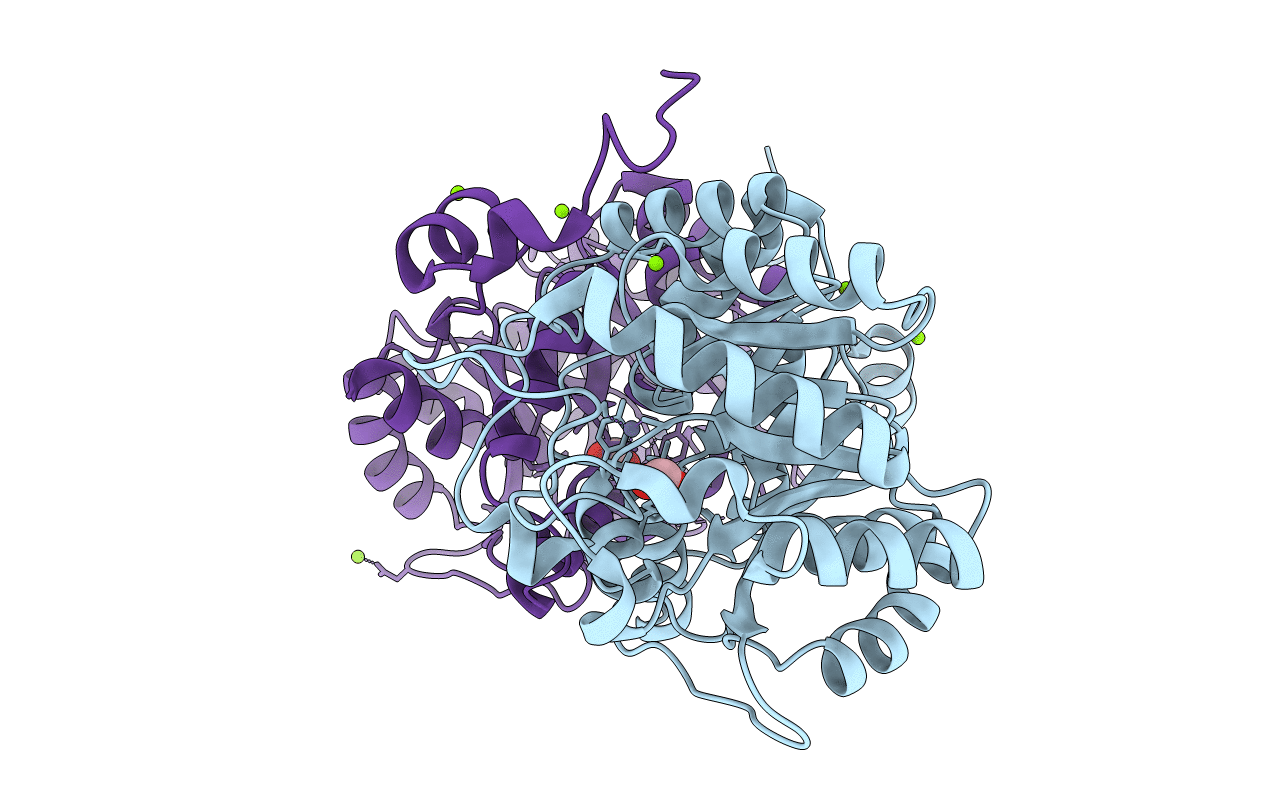
Stepwise introduction of zinc binding site into porphobilinogen synthase of Pseudomonas aeruginosa (mutations A129C and D139C)
Biological Source:
Source Organism:
Host Organism:
Method Details:
Experimental Method:
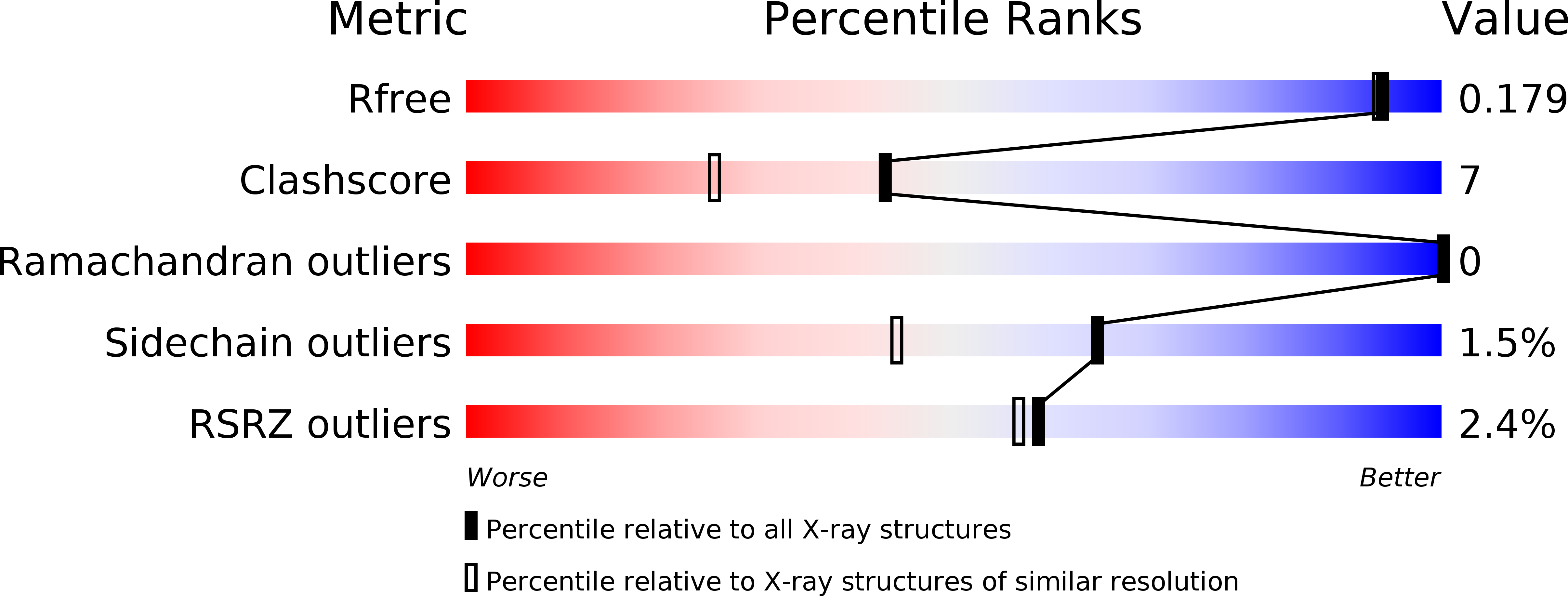
Resolution:
1.60 Å
R-Value Free:
0.16
R-Value Work:
0.12
R-Value Observed:
0.13
Space Group:
P 4 21 2
Macromolecular Entities
Polymer Type:polypeptide(L)
Molecule:DELTA-AMINOLEVULINIC ACID DEHYDRATASE
Gene (Uniprot):hemB
Mutations:YES
Chain IDs:A, B
Chain Length:337
Number of Molecules:2
Biological Source:PSEUDOMONAS AERUGINOSA
Primary Citation
Tracking the Evolution of Porphobilinogen Synthase Metal Dependence in Vitro
J.Mol.Biol. 345 1059 ? (2005)
PMID: 15644204 DOI: 10.1016/J.JMB.2004.10.053

Abstact

Metal ions are indispensable cofactors for chemical catalysis by a plethora of enzymes. Porphobilinogen synthases (PBGSs), which catalyse the second step of tetrapyrrole biosynthesis, are grouped according to their dependence on Zn(2+). Using site-directed mutagenesis, we embarked on transforming Zn(2+)-independent Pseudomonas aeruginosa PBGS into a Zn(2+)-dependent enzyme. Nine PBGS variants were generated by permutationally introducing three cysteine residues and a further two residues into the active site of the enzyme to match the homologous Zn(2+)-containing PBGS from Escherichia coli. Crystal structures of seven enzyme variants were solved to elucidate the nature of Zn(2+) coordination at high resolution. The three single-cysteine variants were invariably found to be enzymatically inactive and only one (D139C) was found to bind detectable amounts of Zn(2+). The double mutant A129C/D139C is enzymatically active and binds Zn(2+) in a tetrahedral coordination. Structurally and functionally it mimics mycobacterial PBGS, which bears an equivalent Zn(2+)-coordination site. The remaining two double mutants, without known natural equivalents, reveal strongly distorted tetrahedral Zn(2+)-binding sites. Variant A129C/D131C possesses weak PBGS activity while D131C/D139C is inactive. The triple mutant A129C/D131C/D139C, finally, displays an almost ideal tetrahedral Zn(2+)-binding geometry and a significant Zn(2+)-dependent enzymatic activity. Two additional amino acid exchanges further optimize the active site architecture towards the E.coli enzyme with an additional increase in activity. Our study delineates the potential evolutionary path between Zn(2+)-free and Zn(2+)-dependent PBGS enyzmes showing that the rigid backbone of PBGS enzymes is an ideal framework to create or eliminate metal dependence through a limited number of amino acid exchanges.

Legend

Protein

Chemical

Disease

Primary Citation of related structures