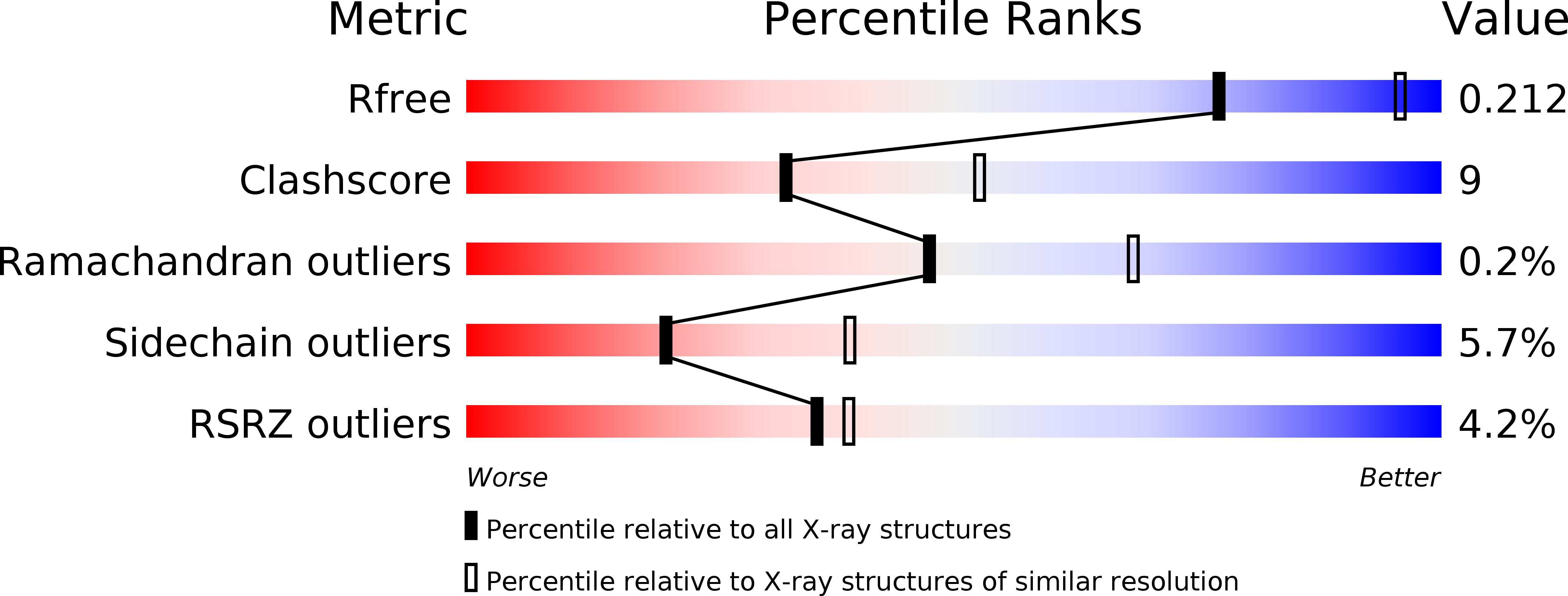
1W4Z image
Deposition Date 2004-08-03
Release Date 2004-12-10
Last Version Date 2023-12-13
Entry Detail
PDB ID:
1W4Z
Title:
Structure of actinorhodin polyketide (actIII) Reductase
Biological Source:
Source Organism:
Host Organism:
Method Details:
Experimental Method:
Resolution:
2.50 Å
R-Value Free:
0.21
R-Value Work:
0.18
R-Value Observed:
0.18
Space Group:
P 32 2 1
Macromolecular Entities
Structures with similar UniProt ID
Protein Blast
Polymer Type:polypeptide(L)
Molecule:KETOACYL REDUCTASE
Gene (Uniprot):actIII
Chain IDs:A, B
Chain Length:281
Number of Molecules:2
Biological Source:STREPTOMYCES COELICOLOR
Primary Citation
The Crystal Structure of the Actiii Actinorhodin Polyketide Reductase; Proposed Mechanism for Acp and Polyketide Binding
Structure 12 1865 ? (2004)
PMID: 15458634 DOI: 10.1016/J.STR.2004.08.002

Abstact

We have determined the 2.5 angstroms crystal structure of an active, tetrameric Streptomyces coelicolor type II polyketide ketoreductase (actIII) with its bound cofactor, NADP+. This structure shows a Rossman dinucleotide binding fold characteristic of SDR enzymes. Of two subunits in the crystallographic asymmetric unit, one is closed around the active site. Formate is observed in the open subunit, indicating possible carbonyl binding sites of the polyketide intermediate. Unlike previous models we observe crystal contacts that may mimic the KR-ACP interactions that may drive active site opening. Based on these observations, we have constructed a model for ACP and polyketide binding. We propose that binding of ACP triggers a conformational change from the closed to the open, active form of the enzyme. The polyketide chain enters the active site and reduction occurs. The model also suggests a general mechanism for ACP recognition which is applicable to a range of protein families.

Legend

Protein

Chemical

Disease

Primary Citation of related structures