1W3F image
Entry Detail
PDB ID:
1W3F
Keywords:
Title:
Crystal structure of the hemolytic lectin from the mushroom Laetiporus sulphureus complexed with N-acetyllactosamine in the gamma motif
Biological Source:
Source Organism:
PDB Version:
Deposition Date:
2004-07-15
Release Date:
2005-02-01
Method Details:
Experimental Method:
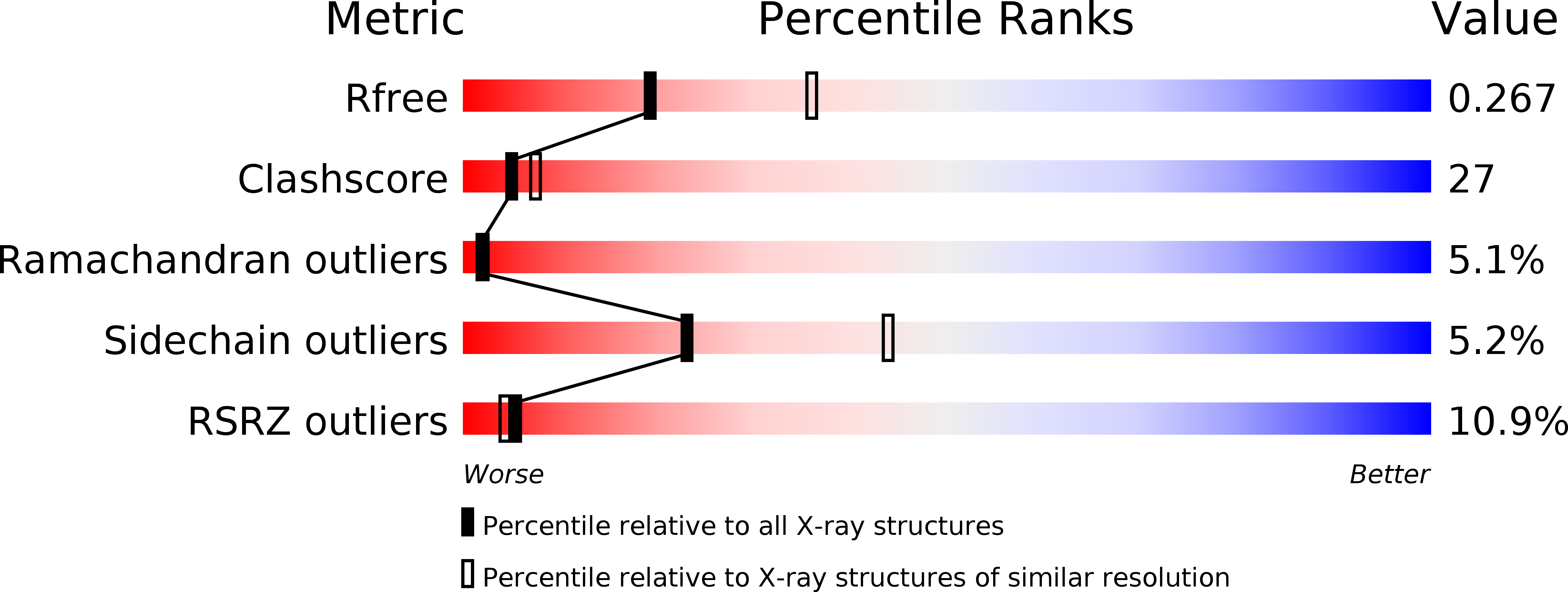
Resolution:
2.58 Å
R-Value Free:
0.27
R-Value Work:
0.23
R-Value Observed:
0.23
Space Group:
P 63 2 2
Macromolecular Entities
Polymer Type:polypeptide(L)
Description:HEMOLYTIC LECTIN FROM LAETIPORUS SULPHUREUS
Chain IDs:A
Chain Length:315
Number of Molecules:1
Biological Source:LAETIPORUS SULPHUREUS
Ligand Molecules
Peptide-like Molecules
PRD_900019
Primary Citation
Structural Analysis of the Laetiporus Sulphureus Hemolytic Pore-Forming Lectin in Complex with Sugars
J.Biol.Chem. 280 17251 ? (2005)
PMID: 15687495 DOI: 10.1074/JBC.M413933200

Abstact

LSL is a lectin produced by the parasitic mushroom Laetiporus sulphureus, which exhibits hemolytic and hemagglutinating activities. Here, we report the crystal structure of LSL refined to 2.6-A resolution determined by the single isomorphous replacement method with the anomalous scatter (SIRAS) signal of a platinum derivative. The structure reveals that LSL is hexameric, which was also shown by analytical ultracentrifugation. The monomeric protein (35 kDa) consists of two distinct modules: an N-terminal lectin module and a pore-forming module. The lectin module has a beta-trefoil scaffold that bears structural similarities to those present in toxins known to interact with galactose-related carbohydrates such as the hemagglutinin component (HA1) of the progenitor toxin from Clostridium botulinum, abrin, and ricin. On the other hand, the C-terminal pore-forming module (composed of domains 2 and 3) exhibits three-dimensional structural resemblances with domains 3 and 4 of the beta-pore-forming toxin aerolysin from the Gram-negative bacterium Aeromonas hydrophila, and domains 2 and 3 from the epsilon-toxin from Clostridium perfringens. This finding reveals the existence of common structural elements within the aerolysin-like family of toxins that could be directly involved in membrane-pore formation. The crystal structures of the complexes of LSL with lactose and N-acetyllactosamine reveal two dissacharide-binding sites per subunit and permits the identification of critical residues involved in sugar binding.

Legend

Protein

Chemical

Disease

Primary Citation of related structures