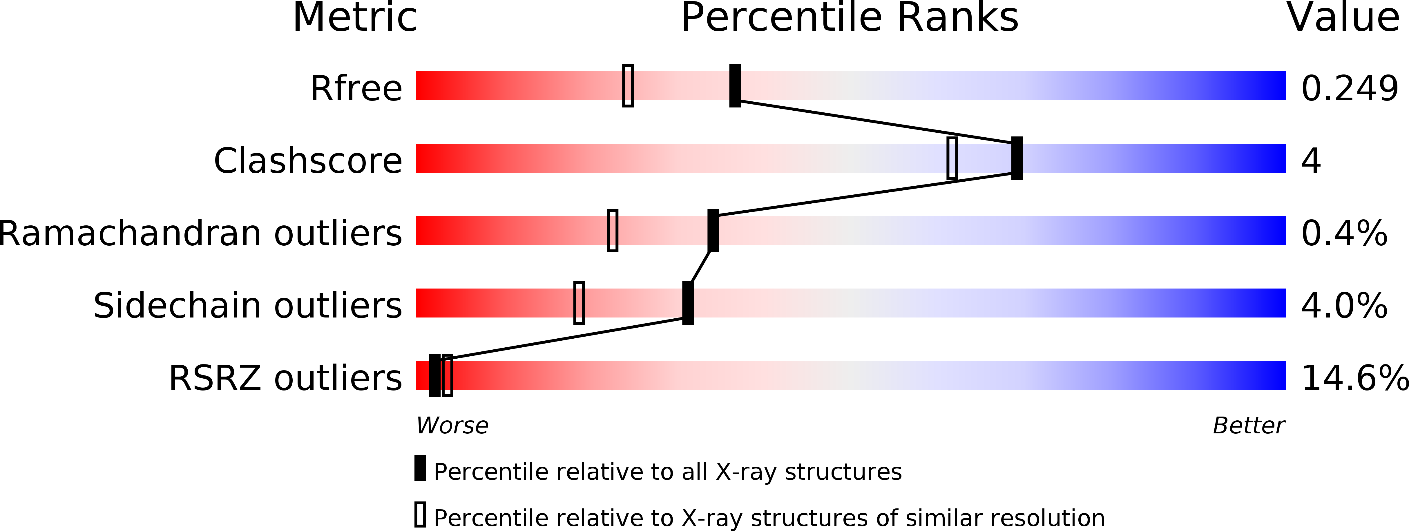
1W2C image
Deposition Date 2004-07-01
Release Date 2004-09-09
Last Version Date 2024-05-08
Entry Detail
PDB ID:
1W2C
Keywords:
Title:
Human Inositol (1,4,5) trisphosphate 3-kinase complexed with Mn2+/AMPPNP/Ins(1,4,5)P3
Biological Source:
Source Organism:
HOMO SAPIENS (Taxon ID: 9606)
Method Details:
Experimental Method:
Resolution:
1.95 Å
R-Value Free:
0.24
R-Value Work:
0.19
R-Value Observed:
0.20
Space Group:
C 2 2 21
Macromolecular Entities
Structures with similar UniProt ID
Protein Blast
Polymer Type:polypeptide(L)
Molecule:INOSITOL-TRISPHOSPHATE 3-KINASE A
Gene (Uniprot):ITPKA
Chain IDs:A, B
Chain Length:265
Number of Molecules:2
Biological Source:HOMO SAPIENS
Primary Citation
Structure of a Human Inositol 1,4,5-Trisphosphate 3-Kinase; Substrate Binding Reveals Why It is not a Phosphoinositide 3-Kinase
Mol.Cell 15 689 ? (2004)
PMID: 15350214 DOI: 10.1016/J.MOLCEL.2004.08.004

Abstact

Mammalian cells produce a variety of inositol phosphates (InsPs), including Ins(1,4,5)P3 that serves both as a second messenger and as a substrate for inositol polyphosphate kinases (IPKs), which further phosphorylate it. We report the structure of an IPK, the human Ins(1,4,5)P3 3-kinase-A, both free and in complexes with substrates and products. This enzyme catalyzes transfer of a phosphate from ATP to the 3-OH of Ins(1,4,5)P3, and its X-ray crystal structure provides a template for understanding a broad family of InsP kinases. The catalytic domain consists of three lobes. The N and C lobes bind ATP and resemble protein and lipid kinases, despite insignificant sequence similarity. The third lobe binds inositol phosphate and is a unique four-helix insertion in the C lobe. This lobe embraces all of the phosphates of Ins(1,4,5)P3 in a positively charged pocket, explaining the enzyme's substrate specificity and its inability to phosphorylate PtdIns(4,5)P2, the membrane-resident analog of Ins(1,4,5)P3.

Legend

Protein

Chemical

Disease

Primary Citation of related structures