1W1I image
Deposition Date 2004-06-22
Release Date 2004-09-02
Last Version Date 2024-11-20
Entry Detail
PDB ID:
1W1I
Keywords:
Title:
Crystal structure of dipeptidyl peptidase IV (DPPIV or CD26) in complex with adenosine deaminase
Biological Source:
Source Organism:
HOMO SAPIENS (Taxon ID: 9606)
BOS TAURUS (Taxon ID: 9913)
Host Organism:
Method Details:
Experimental Method:
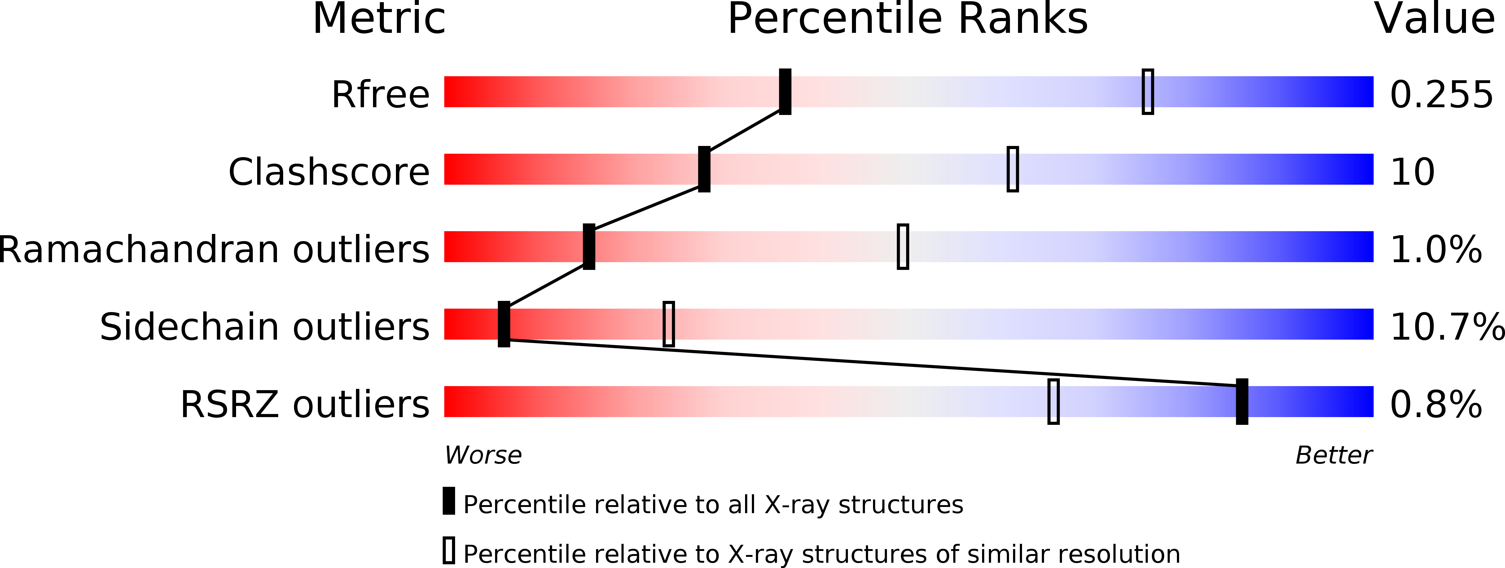
Resolution:
3.03 Å
R-Value Free:
0.25
R-Value Work:
0.22
R-Value Observed:
0.22
Space Group:
C 1 2 1
Macromolecular Entities
Structures with similar UniProt ID
Protein Blast
Polymer Type:polypeptide(L)
Molecule:DIPEPTIDYL PEPTIDASE IV
Gene (Uniprot):DPP4
Chain IDs:A, B, C, D
Chain Length:728
Number of Molecules:4
Biological Source:HOMO SAPIENS
Structures with similar UniProt ID
Protein Blast
Polymer Type:polypeptide(L)
Molecule:ADENOSINE DEAMINASE
Gene (Uniprot):ADA
Chain IDs:E, F, G, H
Chain Length:357
Number of Molecules:4
Biological Source:BOS TAURUS
Modified Residue
Compound ID Chain ID Parent Comp ID Details 2D Image
ASN A ASN GLYCOSYLATION SITE
Primary Citation
Crystal structure of CD26/dipeptidyl-peptidase IV in complex with adenosine deaminase reveals a highly amphiphilic interface.
J. Biol. Chem. 279 43330 43335 (2004)
PMID: 15213224 DOI: 10.1074/jbc.M405001200

Abstact

Dipeptidyl-peptidase IV (DPPIV or CD26) is a homodimeric type II membrane glycoprotein in which the two monomers are subdivided into a beta-propeller domain and an alpha/beta-hydrolase domain. As dipeptidase, DPPIV modulates the activity of various biologically important peptides and, in addition, DPPIV acts as a receptor for adenosine deaminase (ADA), thereby mediating co-stimulatory signals in T-lymphocytes. The 3.0-A resolution crystal structure of the complex formed between human DPPIV and bovine ADA presented here shows that each beta-propeller domain of the DPPIV dimer binds one ADA. At the binding interface, two hydrophobic loops protruding from the beta-propeller domain of DPPIV interact with two hydrophilic and heavily charged alpha-helices of ADA, giving rise to the highest percentage of charged residues involved in a protein-protein contact reported thus far. Additionally, four glycosides linked to Asn229 of DPPIV bind to ADA. In the crystal structure of porcine DPPIV, the observed tetramer formation was suggested to mediate epithelial and lymphocyte cell-cell adhesion. ADA binding to DPPIV could regulate this adhesion, as it would abolish tetramerization.

Legend

Protein

Chemical

Disease

Primary Citation of related structures